Millipore Sigma Vibrant Logo

OP171 Anti-CD63 Mouse mAb (NKI/C-3)

OP171
  
Recuperando precio...
No pudo obtenerse el precio
La cantidad mínima tiene que ser múltiplo de
Maximum Quantity is
Al finalizar el pedido Más información
Ahorró ()
 
Solicitar precio
Disponibilidad a confirmar
Ingrese cantidad 
Suspendido
Cantidades limitadas disponibles
Debe confirmarse disponibilidad
    El resto: se avisará
      El resto: se avisará
      Se avisará
      Póngase en contacto con el Servicio de Atención al Cliente
      Contact Customer Service


      Póngase en contacto con el Servicio de Atención al Cliente

      Descripción

      Replacement Information

      Tabla espec. clave

      Species ReactivityHostAntibody Type
      HMMonoclonal Antibody
      Description
      Overview

      This product has been discontinued.


      Recognizes the heterogeneous 25-110 kDa CD63 glycoprotein in melanomas, clear cell sarcomas, nevocellular nevi, and normal melanocytes. Also reacts with some mucous-producing tumors, carcinoids, carcinomas of the thyroid, mast cells, histiocytes in tumor regions, and cells with secretory function.

      Catalogue NumberOP171
      Brand Family Calbiochem®
      Application Data
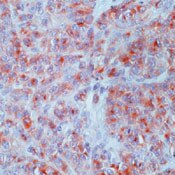
      Detection of human CD63 by immunohistochemistry. Sample: Human melanoma fixed in formalin and paraffin-embedded. Primary antibody: Anti-CD63 Mouse mAb (NKI/C-3) (Cat. No. OP171) (1:500). Detection: fluorescence.
      References
      ReferencesHagen, E.C., et al. 1986. Histopathology 10, 689
      Vennegoor, C., et al. 1986. Cancer Immunol. Immunother. 23, 93.
      Palmer, A.A., et al. 1985. Pathology 17, 335.
      Product Information
      FormLiquid
      FormulationIn 10 mM PBS, 0.2% BSA, pH 7.4.
      Positive controlMelanoma tissue
      Preservative≤0.1% sodium azide
      Applications
      Application ReferencesImmunohistochemistry Stone, CH., et al. 2001. Arch. Pathol. Lab. Med. 125, 751. Bar, H., and Schlote, W. 1997. Clin. Neuropathol. 16, 337. Fernando, S.S., et al. 1994. Pathology 26, 16. Immunoblotting Wang, R., et al. 2001. J. Immunol. Methods 249, 167.
      Key Applications Immunoblotting (Western Blotting)
      Paraffin Sections
      Application NotesImmunoblotting (see application references)
      Paraffin Sections (0.2-0.4 µg/ml, heat pre-treatment required; see application references)
      Application CommentsExhibits cytoplasmic staining when used in immunohistochemistry. Staining of formalin-fixed, paraffin-embedded tissue sections requires boiling in 10 mM citrate buffer, pH 6.0, for 10-20 min, followed by cooling at room temperature for 20 min. Antibody should be titrated for optimal results in individual systems.
      Biological Information
      Immunogenplasma membrane fraction of human MeWo cells
      ImmunogenHuman
      CloneNKI/C-3
      HostMouse
      IsotypeIgG₁
      Species Reactivity
      • Human
      Antibody TypeMonoclonal Antibody
      Concentration Label Please refer to vial label for lot-specific concentration
      Physicochemical Information
      Dimensions
      Materials Information
      Toxicological Information
      Safety Information according to GHS
      Safety Information
      Product Usage Statements
      Storage and Shipping Information
      Ship Code Blue Ice Only
      Toxicity Standard Handling
      Storage +2°C to +8°C
      Do not freeze Yes
      Packaging Information
      Transport Information
      Supplemental Information
      Specifications
      Global Trade Item Number
      Número de referencia GTIN
      OP171 0

      Documentation

      Anti-CD63 Mouse mAb (NKI/C-3) Ficha datos de seguridad (MSDS)

      Título

      Ficha técnica de seguridad del material (MSDS) 

      Anti-CD63 Mouse mAb (NKI/C-3) Certificados de análisis

      CargoNúmero de lote
      OP171

      Referencias bibliográficas

      Visión general referencias
      Hagen, E.C., et al. 1986. Histopathology 10, 689
      Vennegoor, C., et al. 1986. Cancer Immunol. Immunother. 23, 93.
      Palmer, A.A., et al. 1985. Pathology 17, 335.
      Ficha técnica

      Note that this data sheet is not lot-specific and is representative of the current specifications for this product. Please consult the vial label and the certificate of analysis for information on specific lots. Also note that shipping conditions may differ from storage conditions.

      Revision02-October-2007 RFH
      ApplicationImmunoblotting (see application references)
      Paraffin Sections (0.2-0.4 µg/ml, heat pre-treatment required; see application references)
      Application Data
      Detection of human CD63 by immunohistochemistry. Sample: Human melanoma fixed in formalin and paraffin-embedded. Primary antibody: Anti-CD63 Mouse mAb (NKI/C-3) (Cat. No. OP171) (1:500). Detection: fluorescence.
      DescriptionProtein G purified mouse monoclonal antibody generated by immunizing BALB/c mice with the specified immunogen and fusing splenocytes with mouse myeloma P3X62Ag8 cells. Recognizes the heterogeneous ~25-110 kDa CD63 glycoprotein.
      BackgroundCD63 is a glycosyl phosphatidyl inositol (GPI)-anchored, granulocyte-specific activation antigen whose expression is up-regulated in activated neutrophils. It exists as a heterogeneous 25-110 kDa glycoprotein that is located mainly in the innerside of membranes of cytoplasmic vesicles in melanoma cells. The unglycosylated 25 kDa precursor can be detected in tissue sections by immunohistochemistry. CD63 can be detected in melanomas, clear cell sarcomas (melanoma of soft tissue), nevocellular nevi, and normal melanocytes. It has also been detected in some mucus producing tumors, carcinoids, carcinomas of the thyroid, mast cells, histiocytes in tumor regions, and in secretory cells such as salivary glands, bronchial glands, sweat glands, pancreas, and prostate.
      HostMouse
      Immunogen speciesHuman
      Immunogenplasma membrane fraction of human MeWo cells
      CloneNKI/C-3
      IsotypeIgG₁
      Specieshuman
      Positive controlMelanoma tissue
      FormLiquid
      FormulationIn 10 mM PBS, 0.2% BSA, pH 7.4.
      Concentration Label Please refer to vial label for lot-specific concentration
      Preservative≤0.1% sodium azide
      CommentsExhibits cytoplasmic staining when used in immunohistochemistry. Staining of formalin-fixed, paraffin-embedded tissue sections requires boiling in 10 mM citrate buffer, pH 6.0, for 10-20 min, followed by cooling at room temperature for 20 min. Antibody should be titrated for optimal results in individual systems.
      Storage +2°C to +8°C
      Do Not Freeze Yes
      Toxicity Standard Handling
      ReferencesHagen, E.C., et al. 1986. Histopathology 10, 689
      Vennegoor, C., et al. 1986. Cancer Immunol. Immunother. 23, 93.
      Palmer, A.A., et al. 1985. Pathology 17, 335.
      Application referencesImmunohistochemistry Stone, CH., et al. 2001. Arch. Pathol. Lab. Med. 125, 751. Bar, H., and Schlote, W. 1997. Clin. Neuropathol. 16, 337. Fernando, S.S., et al. 1994. Pathology 26, 16. Immunoblotting Wang, R., et al. 2001. J. Immunol. Methods 249, 167.