


ST1705 Sigma-AldrichAnti-TOMM20 Mouse mAb (4F3)
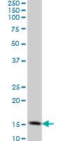

This Anti-TOMM20 Mouse mAb (4F3) is validated for use in ELISA, Immunoblotting, Immunocytochemistry, Paraffin Sections for the detection of TOMM20.
More>> This Anti-TOMM20 Mouse mAb (4F3) is validated for use in ELISA, Immunoblotting, Immunocytochemistry, Paraffin Sections for the detection of TOMM20. Less<<Anti-TOMM20 Mouse mAb (4F3) MSDS (material safety data sheet) or SDS, CoA and CoQ, dossiers, brochures and other available documents.
Synonyms: Anti-Translocase of Outer Mitochondrial Membrane 20
Overview
| Replacement Information |
|---|
Key Spec Table
| Species Reactivity | Host | Antibody Type |
|---|---|---|
| H, M, R | M | Monoclonal Antibody |
Pricing & Availability
| Catalogue Number | Availability | Packaging | Qty/Pack | Price | Quantity | |
|---|---|---|---|---|---|---|
| ST1705-100UG |
|
100 μg |
|
— |


| References |
|---|
| Product Information | |
|---|---|
| Form | liquid |
| Formulation | In PBS, pH 7.2. |
| Negative control | 293T cells |
| Positive control | HeLa cells, PC12 cells, NIH3T3 cells, Human small intestine tissue |
| Preservative | None |
| Quality Level | MQ100 |
| Physicochemical Information |
|---|
| Dimensions |
|---|
| Materials Information |
|---|
| Toxicological Information |
|---|
| Safety Information according to GHS |
|---|
| Safety Information |
|---|
| Product Usage Statements |
|---|
| Packaging Information |
|---|
| Transport Information |
|---|
| Supplemental Information |
|---|
| Specifications |
|---|
| Global Trade Item Number | |
|---|---|
| Catalogue Number | GTIN |
| ST1705-100UG | 04055977207347 |
Documentation
Anti-TOMM20 Mouse mAb (4F3) SDS
| Title |
|---|