


ST1204 Sigma-AldrichAnti-PGC-1α Rabbit pAb

This Anti-PGC-1α Rabbit pAb is validated for use in Immunoblotting, Immunoprecipitation for the detection of PGC-1α.
More>> This Anti-PGC-1α Rabbit pAb is validated for use in Immunoblotting, Immunoprecipitation for the detection of PGC-1α. Less<<Anti-PGC-1α Rabbit pAb MSDS (material safety data sheet) or SDS, CoA and CoQ, dossiers, brochures and other available documents.
Recommended Products
Overview
| Replacement Information |
|---|
Key Spec Table
| Species Reactivity | Host | Antibody Type |
|---|---|---|
| H, M, R | Rb | Polyclonal Antibody |
Pricing & Availability
| Catalogue Number | Availability | Packaging | Qty/Pack | Price | Quantity | |
|---|---|---|---|---|---|---|
| ST1204-100UL |
|
100 ul |
|
— |

| Product Information | |
|---|---|
| Form | Liquid |
| Formulation | In 50 mM PBS |
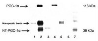
| Positive control | Brown adipose tissue from mice exposed to cold |
| Preservative | None |
| Quality Level | MQ100 |
| Physicochemical Information |
|---|
| Dimensions |
|---|
| Materials Information |
|---|
| Toxicological Information |
|---|
| Safety Information according to GHS |
|---|
| Safety Information |
|---|
| Product Usage Statements |
|---|
| Packaging Information |
|---|
| Transport Information |
|---|
| Supplemental Information |
|---|
| Specifications |
|---|
| Global Trade Item Number | |
|---|---|
| Catalogue Number | GTIN |
| ST1204-100UL | 04055977224108 |
Documentation
Anti-PGC-1α Rabbit pAb SDS
| Title |
|---|
Anti-PGC-1α Rabbit pAb Certificates of Analysis
| Title | Lot Number |
|---|---|
| ST1204 |
References
| Reference overview |
|---|
| Chang, J.S., et al. 2010. J. Biol. Chem. 285, 18039. Zhang, Y., et al. 2009. J. Biol. Chem. 284, 32813. Lai, L., et al. 2008. Genes Dev. 14, 1948. Rodgers, J.T., et al. 2008. FEBS Lett. 582, 46. Mazzucotelli, A., et al. 2007. Diabetes 10, 2467. Nemoto, S., et al. 2005. J. Biol. Chem. 16, 16456. |