Our broad portfolio consists of multiplex panels that allow you to choose, within the panel, analytes that best meet your needs. On a separate tab you can choose the premixed cytokine format or a single plex kit.
Cell Signaling Kits & MAPmates™
Choose fixed kits that allow you to explore entire pathways or processes. Or design your own kits by choosing single plex MAPmates™, following the provided guidelines.
The following MAPmates™ should not be plexed together:
-MAPmates™ that require a different assay buffer
-Phospho-specific and total MAPmate™ pairs, e.g. total GSK3β and GSK3β (Ser 9)
-PanTyr and site-specific MAPmates™, e.g. Phospho-EGF Receptor and phospho-STAT1 (Tyr701)
-More than 1 phospho-MAPmate™ for a single target (Akt, STAT3)
-GAPDH and β-Tubulin cannot be plexed with kits or MAPmates™ containing panTyr
.
Catalogue Number
Ordering Description
Qty/Pack
List
This item has been added to favorites.
Select A Species, Panel Type, Kit or Sample Type
To begin designing your MILLIPLEX® MAP kit select a species, a panel type or kit of interest.
Custom Premix Selecting "Custom Premix" option means that all of the beads you have chosen will be premixed in manufacturing before the kit is sent to you.
Catalogue Number
Ordering Description
Qty/Pack
List
This item has been added to favorites.
Species
Panel Type
Selected Kit
Qty
Catalogue Number
Ordering Description
Qty/Pack
List Price
96-Well Plate
Qty
Catalogue Number
Ordering Description
Qty/Pack
List Price
Add Additional Reagents (Buffer and Detection Kit is required for use with MAPmates)
Qty
Catalogue Number
Ordering Description
Qty/Pack
List Price
48-602MAG
Buffer Detection Kit for Magnetic Beads
1 Kit
Space Saver Option Customers purchasing multiple kits may choose to save storage space by eliminating the kit packaging and receiving their multiplex assay components in plastic bags for more compact storage.
This item has been added to favorites.
The Product Has Been Added To Your Cart
You can now customize another kit, choose a premixed kit, check out or close the ordering tool.
IM35
Sigma-AldrichAnti-MMP-1 (Ab-1) Mouse mAb (41-1E5)
The quantity field is empty. Please enter a quantity of 1 or more to add items to your cart.
Description
Overview
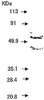
Recognizes the ~55 kDa latent and the ~43 kDa active forms of MMP-1.
Catalogue Number
IM35
Brand Family
Calbiochem®
Application Data
Detection of human MMP-1 by immunoblotting. Sample: Conditioned medium of normal human skin fibroblasts CCD-41SK cells. Primary antibody: Anti-MMP-1 (Ab-1) Mouse mAb (41-1E5) (Cat. No. IM35) (1 µg/ml). Detection: chemiluminescence.
References
References
Cottam, D.W. and Rees, R.C. 1993. Intl. J. Oncol.2, 861. Stetler-Stevenson, W.G., et al. 1993. FASEB7, 1434. Zhang, J., et al. 1993. Clinica Chimica Acta.219, 1. Woessner, J.F. 1991. FASEB5, 2145. Liotta, L.A. and Stetler-Stevenson, W.G. 1990. in Seminars in Cancer Biology, ed. M. M. Gottesman. Vol. 1; 99-106.
Product Information
Declaration
Manufactured by Daiichi Fine Chemical Co., Ltd. Not available for sale in Japan.
Form
Liquid
Formulation
In 100 mM sodium phosphate buffer, 0.1% BSA, pH 7.0
To prepare conditioned medium for positive control, incubate HT-1080 cells for 2 h at 37°C in serum-free media containing 100 nM PMA. Collect and centrifuge medium; concentrate as necessary. Does not cross-react with bovine. Will not cross-react with human MMP-2, MMP-3, MMP-8, MMP-9, or MMP-13. For best results with paraffin sections, pre-treat with a pressure cooker; however, heat, saponin or trypsin can be used in place of a pressure cooker. Antibody should be titrated for optimal results in individual systems.
Biological Information
Immunogen
a synthetic peptide (VQGQNVLHGYPKDIYSSFG) corresponding to amino acids 332-350 of human MMP-1
Immunogen
Human
Clone
41-1E5
Host
Mouse
Isotype
IgG2a
Species Reactivity
Human
Antibody Type
Monoclonal Antibody
Concentration Label
Please refer to vial label for lot-specific concentration
Storage and Shipping Information
Ship Code
Blue Ice Only
Toxicity
Standard Handling
Storage
-20°C
Avoid freeze/thaw
Avoid freeze/thaw
Do not freeze
Ok to freeze
Special Instructions
Following initial thaw, aliquot and freeze (-20°C).
Anti-MMP-1 (Ab-1) Mouse mAb (41-1E5) Certificates of Analysis
Title
Lot Number
IM35
References
Reference overview
Cottam, D.W. and Rees, R.C. 1993. Intl. J. Oncol.2, 861. Stetler-Stevenson, W.G., et al. 1993. FASEB7, 1434. Zhang, J., et al. 1993. Clinica Chimica Acta.219, 1. Woessner, J.F. 1991. FASEB5, 2145. Liotta, L.A. and Stetler-Stevenson, W.G. 1990. in Seminars in Cancer Biology, ed. M. M. Gottesman. Vol. 1; 99-106.
Data Sheet
Note that this data sheet is not lot-specific and is representative of the current specifications for this product. Please consult the vial label and the certificate of analysis for information on specific lots. Also note that shipping conditions may differ from storage conditions.
Detection of human MMP-1 by immunoblotting. Sample: Conditioned medium of normal human skin fibroblasts CCD-41SK cells. Primary antibody: Anti-MMP-1 (Ab-1) Mouse mAb (41-1E5) (Cat. No. IM35) (1 µg/ml). Detection: chemiluminescence.
Description
Purified mouse monoclonal antibody. Recognizes the ~55 kDa latent and the ~43 kDa active forms of MMP-1.
Background
Matrix metalloproteinases (MMPs) are a family of enzymes that are responsible for the degradation of extracellular matrix components such as collagen, laminin and proteoglycans. In addition to sequence homology, all MMPs share the following characteristics: the catalytic mechanism is dependent upon a zinc ion at the active center, they cleave one or more extracellular matrix components, they are secreted as zymogens which are activated by removal of an ~10 kDa segment from the N-terminus and they are inhibited by tissue inhibitor of metalloproteinases (TIMP). These enzymes are involved in normal physiological processes such as embryogenesis and tissue remodeling and may play an important role in arthritis, periodontitis, and metastasis.
MMP-1 (interstitial collagenase, tissue collagenase, fibroblast collagenase) is secreted as a 57/52 kDa zymogen which is proteolytically processed to the 46/42 kDa active forms. This enzyme displays substrate specificity toward type I, II, III, VII, VIII and X collagens and gelatin. MMP-1 is thought to play an important role in pathophysiological degradation processes associated with conditions such as rheumatoid arthritis, osteoarthritis, and cancer cell invasion.
Host
Mouse
Immunogen species
Human
Immunogen
a synthetic peptide (VQGQNVLHGYPKDIYSSFG) corresponding to amino acids 332-350 of human MMP-1
Clone
41-1E5
Isotype
IgG2a
Species
not bovine, human
Form
Liquid
Formulation
In 100 mM sodium phosphate buffer, 0.1% BSA, pH 7.0
Concentration Label
Please refer to vial label for lot-specific concentration
Preservative
≤0.1% sodium azide
Comments
To prepare conditioned medium for positive control, incubate HT-1080 cells for 2 h at 37°C in serum-free media containing 100 nM PMA. Collect and centrifuge medium; concentrate as necessary. Does not cross-react with bovine. Will not cross-react with human MMP-2, MMP-3, MMP-8, MMP-9, or MMP-13. For best results with paraffin sections, pre-treat with a pressure cooker; however, heat, saponin or trypsin can be used in place of a pressure cooker. Antibody should be titrated for optimal results in individual systems.
Storage
Avoid freeze/thaw -20°C
Do Not Freeze
Ok to freeze
Special Instructions
Following initial thaw, aliquot and freeze (-20°C).
Toxicity
Standard Handling
References
Cottam, D.W. and Rees, R.C. 1993. Intl. J. Oncol.2, 861. Stetler-Stevenson, W.G., et al. 1993. FASEB7, 1434. Zhang, J., et al. 1993. Clinica Chimica Acta.219, 1. Woessner, J.F. 1991. FASEB5, 2145. Liotta, L.A. and Stetler-Stevenson, W.G. 1990. in Seminars in Cancer Biology, ed. M. M. Gottesman. Vol. 1; 99-106.
Application references
Paraffin Sections
Zhang, J., et al. 1993. Clinica Chimica Acta.219, 1.
Frozen Sections
Nakagawa, T., et al. 1994. J. Neurosurg.81, 69.