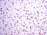
Phenotypic diversity and expression of GABAergic inhibitory interneurons during postnatal development in lumbar spinal cord of glutamic acid decarboxylase 67-green fluorescent protein mice.
Dougherty, KJ; Sawchuk, MA; Hochman, S
Neuroscience
163
909-19
2009
Show Abstract
The synthesis enzyme glutamic acid decarboxylase (GAD65 or GAD67) identifies neurons as GABAergic. Recent studies have characterized the physiological properties of spinal cord GABAergic interneurons using lines of GAD67-green fluorescent protein (GFP) transgenic mice. A more complete characterization of their phenotype is required to better understand the role of this population of inhibitory neurons in spinal cord function. Here, we characterize the distribution of lumbar spinal cord GAD67-GFP neurons at postnatal days (P) 0, 7, and 14, and adult based on their co-expression with GABA and determine the molecular phenotype of GAD67-GFP neurons at P14 based on the expression of various neuropeptides, calcium binding proteins, and other markers. At all ages greater than 67% of GFP(+) neurons were also GABA(+). With increasing age; (i) GFP(+) and GABA(+) cell numbers declined, (ii) ventral horn GFP(+) and GABA(+) neurons vanished, and (iii) somatic labeling was reduced while terminal labeling increased. At P14, vasoactive intestinal peptide and bombesin were expressed in approximately 63% and approximately 35% of GFP(+) cells, respectively. Somatostatin was found in a small number of neurons, whereas calcitonin gene-related peptide never co-localized with GFP. Moderate co-expression was found for all the Ca(2+) binding proteins examined. Notably, most laminae I-II parvalbumin(+) neurons were also GFP(+). Neurogranin, a protein kinase C substrate, was found in approximately 1/2 of GFP(+) cells. Lastly, while only 7% of GFP(+) cells contain nitric oxide synthase (NOS), these cells represent a large fraction of all NOS(+) cells. We conclude that GAD67-GFP neurons represent the majority of spinal GABAergic neurons and that mouse dorsal horn GAD67-GFP(+) neurons comprise a phenotypically diverse population. Full Text Article | 19560523
 |
The presence and absence of prosencephalic cell groups relaying striatal information to the medial and lateral thalamus in tenrec.
Heinz Künzle
Journal of anatomy
212
2008
Show Abstract
Although there are remarkable differences regarding the output organization of basal ganglia between mammals and non-mammals, mammalian species with poorly differentiated brain have scarcely been investigated in this respect. The aim of the present study was to identify the pallidal neurons giving rise to thalamic projections in the Madagascar lesser hedgehog tenrec (Afrotheria). Following tracer injections into the thalamus, retrogradely labelled neurons were found in the depth of the olfactory tubercle (particularly the hilus of the Callejal islands and the insula magna), in subdivisions of the diagonal band complex, the peripeduncular region and the thalamic reticular nucleus. No labelled cells were seen in the globus pallidus. Pallidal neurons were tentatively identified on the basis of their striatal afferents revealed hodologically using anterograde axonal tracer substances and immunohistochemically with antibodies against enkephalin and substance P. The data showed that the tenrec's medial thalamus received prominent projections from ventral pallidal cells as well as from a few neurons within and ventral to the cerebral peduncle. The only regions projecting to the lateral thalamus appeared to be the thalamic reticular nucleus (RTh) and the dorsal peripeduncular nucleus (PpD). On the basis of immunohistochemical data and the topography of its thalamic projections, the PpD was considered to be an equivalent to the pregeniculate nucleus in other mammals. There was no evidence of entopeduncular (internal pallidal) neurons being present within the RTh/PpD complex, neuropils of which did not stain for enkephalin and substance P. The ventrolateral portion of RTh, the only region eventually receiving a striatal input, projected to the caudolateral rather than the rostrolateral thalamus. Thus, the striatopallidal output organization in the tenrec appeared similar, in many respects, to the output organization in non-mammals. This paper considers the failure to identify entopeduncular neurons projecting to the rostrolateral thalamus in a mammal with a little differentiated cerebral cortex, and also stresses the discrepancy between this absence and the presence of a distinct external pallidal segment (globus pallidus). Full Text Article | 18510507
 |


