Millipore Sigma Vibrant Logo

CBA047 Active Caspase-9 Assay Kit

CBA047
  
Retrieving price...
Price could not be retrieved
Minimum Quantity is a multiple of
Maximum Quantity is
Upon Order Completion More Information
You Saved ()
 
Request Pricing
Limited Availability
In Stock 
Discontinued
Limited Quantities Available
Availability to be confirmed
    Remaining : Will advise
      Remaining : Will advise
      Will advise
      Contact Customer Service
      Contact Customer Service


      Contact Customer Service

      Overview

      Replacement Information
      Description
      OverviewCaspases belong to a family of cysteine proteases that cleave substrates at the carboxyl terminus of asparate residues. Caspase-9 is an initiator caspase responsible for activation of the intrinsic pathway of apoptosis. This is a convenient immunofluorescent assay for the quantitative detection of active caspase-9 in cell lysates.
      Catalogue NumberCBA047
      Brand Family Calbiochem®
      Materials Required but Not DeliveredFilter all buffers through a 0.2 m filter and store at 4°C unless noted otherwise. PBS - 137 mM NaCl, 2.7 mM KCl, 8.1 mM NaH2PO4, 1.5 mM KH2PO4, pH to 7.2-7.4. Wash Buffer - PBS containing 0.05% Tween®-20 detergent. Make fresh before each use. Block Buffer - PBS containing 5% sucrose and 1% bovine serum albumin (BSA). Make fresh before each use. Assay Buffer - 100 mM KCl, 1 mM EGTA, 1 mM EDTA, 1 mM MgCl2, 5 mM DTT, 5% PEG (polyethylene glycol), 0.1% CHAPS, and 50 mM HEPES. pH to 7.5 with KOH. Caspase-9 Capture Buffer - 50 mM KCl, 5 mM EGTA, 1 mM MgCl2, 5 mM DTT, 5% sucrose, and 50 mM HEPES. pH to 7.5 with KOH. Caspase-9 Lysis Buffer 1-50 mM KCl, 5 mM EGTA, 2 mM MgCl2, 1 mM DTT, 0.2% CHAPS, and 50 mM HEPES. pH to 7.5 with KOH. Caspase-9 Lysis Buffer 2-50 mM KCl, 5 mM EGTA, 1 mM MgCl2, 5 mM DTT, 0.2% CHAPS, 5% sucrose, and 50 mM HEPES. pH to 7.5 with KOH. Bovine cytochrome c - Prepare a 2 mg/ml solution in Caspase-9 Lysis Buffer 1 without protease inhibitors and store at -20°C in a manual defrost freezer for up to 1 week. Avoid repeated freeze-thaw cycles. dATP - Prepare a 10 mM solution in Caspase-9 Lysis Buffer 1 without protease inhibitors and adjust pH to 7.5 with KOH. Aliquot and store at -20°C in a manual defrost freezer for up to 1 week. Avoid repeated freeze-thaw cycles. Cytochalasin B (Cat. No. 250233) Chymostatin (Cat. No. 230790) Leupeptin (Cat. No. 108975) Antipain (Cat. No. 178223) Pepstatin (Cat. No. 516481) Phenylmethylsulfonylfluoride (PMSF) (Cat. No. 52332) Polyethylene Glycol (PEG 8000) DMSO KOH Pipettes and pipette tips Deionized or distilled water 96-well plates Plate sealers Multi-channel pipette, squirt bottle, manifold dispenser, or automated plate washer 30°C and 37°C water bath or incubator Fluorescent plate reader capable of measuring the kinetics of enzymatic reactions at 400nm excitation and 505nm emission wavelengths Centrifuge capable of reaching 12,000 x g
      References
      Product Information
      Detection methodFluorometric
      Form480 Tests
      Format96-well plate
      Kit containsCaspase-9 Capture Antibody, Precursor Caspase-3, DEVD-AFC, and a user protocol.
      Applications
      Biological Information
      Sample TypeCells
      Physicochemical Information
      Dimensions
      Materials Information
      Toxicological Information
      Safety Information according to GHS
      Safety Information
      Product Usage Statements
      Storage and Shipping Information
      Ship Code Blue Ice Only
      Toxicity Standard Handling
      Storage -20°C
      Do not freeze Ok to freeze
      Packaging Information
      Transport Information
      Supplemental Information
      Kit containsCaspase-9 Capture Antibody, Precursor Caspase-3, DEVD-AFC, and a user protocol.
      Specifications
      Global Trade Item Number
      Catalogue Number GTIN
      CBA047 0

      Documentation

      Brochure

      Title
      Kit SourceBook - 2nd Edition EURO
      Kits SourceBook - 2nd Edition GBP
      User Protocol

      Revision08-October-2008 JSW
      Form480 Tests
      Format96-well plate
      Detection methodFluorometric
      Specieshuman
      Principles of the assayThe Calbiochem® Active Caspase-9 Assay Kit captures active caspase-9 complexed with APAF-1. Unbound material is removed by washing and inactive precursor caspase-3, a natural substrate for caspase-9, is added. Active caspase-9 cleaves precursor caspase-3 thereby activating caspase-3. The synthetic peptide substrate DEVD-AFC is added and is cleaved by caspase-3 to generate fluorescent AFC. Fluorescence is measured in a 96-well fluorescent plate reader. Each kit contains sufficient materials to run ELISAs on approximately five 96-well plates, provided that the reagents are prepared as described in this package insert and the assay is run as described in the Detailed Protocol using the recommended plates, buffers, diluents, substrates, and solutions.
      Materials provided• Caspase-9 Capture Antibody (Kit Component No. JA9146): 1 vial • Precursor Caspase-3 (Kit Component No. JA9147): 1 vial • DEVD-AFC (Asp-Glu-Val-Asp 7-Amino-4-Trifluoromethyl Coumarin) (Kit Component No. JA9145): 1 vial, 2 mg, contains lyophilized substrate.
      Materials Required but not providedFilter all buffers through a 0.2 m filter and store at 4°C unless noted otherwise. PBS - 137 mM NaCl, 2.7 mM KCl, 8.1 mM NaH2PO4, 1.5 mM KH2PO4, pH to 7.2-7.4. Wash Buffer - PBS containing 0.05% Tween®-20 detergent. Make fresh before each use. Block Buffer - PBS containing 5% sucrose and 1% bovine serum albumin (BSA). Make fresh before each use. Assay Buffer - 100 mM KCl, 1 mM EGTA, 1 mM EDTA, 1 mM MgCl2, 5 mM DTT, 5% PEG (polyethylene glycol), 0.1% CHAPS, and 50 mM HEPES. pH to 7.5 with KOH. Caspase-9 Capture Buffer - 50 mM KCl, 5 mM EGTA, 1 mM MgCl2, 5 mM DTT, 5% sucrose, and 50 mM HEPES. pH to 7.5 with KOH. Caspase-9 Lysis Buffer 1-50 mM KCl, 5 mM EGTA, 2 mM MgCl2, 1 mM DTT, 0.2% CHAPS, and 50 mM HEPES. pH to 7.5 with KOH. Caspase-9 Lysis Buffer 2-50 mM KCl, 5 mM EGTA, 1 mM MgCl2, 5 mM DTT, 0.2% CHAPS, 5% sucrose, and 50 mM HEPES. pH to 7.5 with KOH. Bovine cytochrome c - Prepare a 2 mg/ml solution in Caspase-9 Lysis Buffer 1 without protease inhibitors and store at -20°C in a manual defrost freezer for up to 1 week. Avoid repeated freeze-thaw cycles. dATP - Prepare a 10 mM solution in Caspase-9 Lysis Buffer 1 without protease inhibitors and adjust pH to 7.5 with KOH. Aliquot and store at -20°C in a manual defrost freezer for up to 1 week. Avoid repeated freeze-thaw cycles. Cytochalasin B (Cat. No. 250233) Chymostatin (Cat. No. 230790) Leupeptin (Cat. No. 108975) Antipain (Cat. No. 178223) Pepstatin (Cat. No. 516481) Phenylmethylsulfonylfluoride (PMSF) (Cat. No. 52332) Polyethylene Glycol (PEG 8000) DMSO KOH Pipettes and pipette tips Deionized or distilled water 96-well plates Plate sealers Multi-channel pipette, squirt bottle, manifold dispenser, or automated plate washer 30°C and 37°C water bath or incubator Fluorescent plate reader capable of measuring the kinetics of enzymatic reactions at 400nm excitation and 505nm emission wavelengths Centrifuge capable of reaching 12,000 x g
      Precautions and recommendations Keep all solutions and samples on ice while performing the assay. Concentrations of enzyme, substrate, and capture antibody used can be varied to create an immunoassay with a different sensitivity and dynamic range. A basic understanding of immunoassay development is required for the successful use of these reagents in immunoassays. A thorough and consistent wash technique is essential for proper assay performance. Wash Buffer should be dispensed forcefully and removed completely from the wells by aspiration or decanting. Remove any remaining Wash Buffer by inverting the plate and blotting it against clean paper towels. Use a fresh reagent reservoir and pipette tips for each step. It is recommended that all samples be assayed in duplicate. Avoid microbial contamination of reagents and buffers. This may interfere with the sensitivity of the assay. Buffers containing protein should be made under sterile conditions and stored at 4°C or be prepared fresh daily.
      Preparation

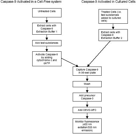
      Figure 1: Sample Preparation

      Caspase-9 may be activated either by adding cytochrome c and dATP to a cell lysate or by adding the appropriate reagents (e.g. staurosporine) to cultured cells.

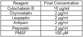
      I. Caspase-9 Activated in a Cell-Free System Using Cell Lysates A. Preparation of Cell Lysates Immediately before harvesting cells, add the following to Caspase-9 Lysis Buffer 1. Place in an ice bath and keep cold.

      Table 1: Preparation of Cell Lysates

      1. Centrifuge cells at 1000 x g for 10 min. Note: If adherent cells are used, scrape cells off the culture flask prior to centrifugation. Save the culture media and PBS wash (step 3), centrifuge, and combine these cells with the cells from step 4. 2. Decant off the culture media, noting the volume. 3. Resuspend cells in a volume of PBS equal to the media volume noted in step 2. 4. Centrifuge cells at 1000 x g for 10 min and remove all PBS. 5. Solubilize cells at 2 x 10⁸ cells/ml in ice cold Caspase-9 Lysis Buffer 1 containing cytochalasin B and protease inhibitors. This high cell concentration is required for activation of caspase-9 in cell lysates. 6. Thoroughly suspend cells by gently pipetting up and down. Incubate on ice for 10 min. 7. Aliquot 225 µl into a 2 ml screw cap microcentrifuge tubes and snap freeze in liquid nitrogen. Note: Snap freezing is essential for complete cell lysis. 8. Store lysate at ≤-70°C or thaw at 4°C and use immediately. Stored samples have been used successfully for up to 1 month after preparation. B. Caspase-9 Activation Please Note: Activation of caspase-9 in cell lystes by addition of cytochrome c and dATP does NOT work with Caspase-9 Lysis Buffer 2 (used in Section II.A.). Thaw stored lysates at 4°C. Activate caspase-9 within 30 min of thawing stored lysates. Perform the assay within 60 min of activation. If multiple peptides/chemical compounds are being tested, the amount of lysate, cytochrome c, and dATP will need to be adjusted. Activated cell lysates in Caspase-9 Lysis Buffer 1 must be diluted a minimum of 4-fold in Caspase-9 Capture Buffer to avoid buffer interference in the assay. The correct dilution will need to be empirically determined. The following protocol is for the preparation of a sufficient amount of activated cell lysate for 5-10 wells of a 96-well plate. 1. Centrifuge 225 µl thawed lysate at 12,000 x g at 4°C for 5 min. 2. Transfer 20 µl of the supernatant to a chilled microcentrifuge tube. Note: If the effects of additional reagents (e.g. IAP inhibitors) are being studied, they should be added at this time. Reduce the volume of the supernatant in step 2, so the final volume remains at 25 µl after addition of cytochrome c and dATP. Adjust controls accordingly. 3. To activate caspase-9 in cell lysates, add 2.5 µl of 2 mg/ml bovine cytochrome c and 2.5 µl of 10 mM dATP. Substitute 5 µl of Caspase-9 Lysis Buffer 1 for cytochrome c and dATP to obtain control lysates. 4. Incubate at 30°C in a water bath for 60 min. 5. Monitor Caspase-9 activation in cell lysates by measuring DEVD-AFC cleavage as follows: Add 2 µl of activated lysates to 88 µl of Assay Buffer. Add 10 µl of 500 µM DEVD-AFC. Immediately monitor fluorescence at 400 nm excitation and 505 nm emission wavelengths using a fluorescent plate reader capable of measuring the kinetics of enzymatic reactions. If no DEVD-AFC cleavage is observed, do not proceed with this lysate or the assay. Begin the protocol again at Step I.B.1. 6. Dilute the activated lysates with Caspase-9 Capture Buffer to obtain an lysate concentration of ~5 x 10⁶ cells/ml. II. Caspase-9 Activated in Cultured Cells A. Preparation of Cell Lysates Immediately before harvesting treated cells, add the following to Caspase-9 Lysis Buffer 2. Place in an ice bath and keep cold.

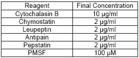
      Table 2: Preparation of Cell Lysates

      1. Centrifuge cells at 1000 x g for 10 min. Note: If adherent cells are used, scrape cells off the flask prior to centrifugation. Certain treatments may cause adherent cells to lift off the flask. In this case, save the culture media and PBS wash (step 3), centrifuge, and combine these cells with those from step 4. 2. Decant off the culture media, noting the volume. 3. Wash cells in a volume of PBS equal to the media volume noted in step 2. 4. Centrifuge cells at 1000 x g for 10 min and remove all PBS. 5. Solubilize cells at a minimum cell concentration of 1 x 10⁷ cells/ml in ice cold Caspase-9 Lysis Buffer 2 containing cytochalasin B and protease inhibitors (Table 1). 6. Thoroughly suspend cells by gently pipetting up and down and incubate on ice for 10 min. 7. Aliquot 225 µl into a 2 ml screw cap microcentrifuge tubes and snap freeze in liquid nitrogen. Note: Snap freezing is essential for complete cell lysis. 8. Store extracts at ≤-70°C or thaw at 4°C and use immediately. Stored samples have been used successfully for up to 1 month after preparation. 9. Centrifuge thawed lysates at 12,000 x g at 4°C for 5 min. 10. Continue with the Detailed Protocol below.
      Reagent preparation• Caspase-9 Capture Antibody: Reconstituted with 1 ml PBS to yield a stock concentration of 360 µg/ml. After reconstitution, store at 4°C for up to 60 days or aliquot and store for up to 3 months at -20°C in a manual defrost freezer or at -70°C. • Precursor Caspase-3: Reconstitute Precursor Caspase-3 in 1 ml Assay Buffer. Aliquot on ice and store at -20°C in a manual defrost freezer. Note: Never allow Precursor Caspase-3 to reach temperatures above 4°C prior to addition to plate wells to prevent autoactivation. • DEVD-AFC (Asp-Glu-Val-Asp 7-Amino-4-Trifluoromethyl Coumarin): Dissolve in 137 µl of DMSO to obtain a 20 mM stock solution. Store at -20°C in a manual defrost freezer. Warm to room temperature and mix thoroughly prior to use. • Plate preparation 1. Dilute the reconstituted Capture Antibody to 6 µg/ml in PBS and add 100 µl per well in a 96-well plate. Seal the plate and incubate overnight at room temperature. 2. Aspirate each well and wash with Wash Buffer, repeating the process two times for a total of 3 washes. Wash by filling each well with Wash Buffer (400 µl) using a squirt bottle, multi-channel pipette, manifold dispenser or autowasher. Complete removal of liquid at each step is essential for good performance. After the last wash, remove any remaining Wash Buffer by aspirating or decanting. Invert the plate and blot it against clean paper towels. 3. Block plates by adding 300 µl Block Buffer to each well. Incubate at room temperature for a minimum of 1 h. 4. Repeat the aspiration/wash as in step 2. The plates are now ready for sample addition. Alternatively, the Block Buffer can be aspirated after step 3 and the plates can be dried under vacuum. When sealed with desiccant, the plates can be stored at 4°C for at least 2 months
      Detailed protocolKeep all solutions and samples on ice while performing this assay. Control Preparation: The following controls should be assayed at the same time as experimental samples. Background levels of DEVD-AFC cleavage and fluorescence are determined by performing the assay with inactive and active lysates with and without Precursor Caspase-3.

      Table 3: Control Preparation

      *Untreated and treated refer to whether or not test peptides or chemical compounds were added to the assay.

      1. Add 100 µl of activated cell lysate from either Step I.B.6 or Step II.A.10 to each well (except those containing controls) of a 96-well plate coated with Capture Antibody. 2. Cover and incubate the plate at room temperature for 2 h. 3. Aspirate samples and wash plates with Caspase-9 Capture Buffer, repeating the process two times for a total of 3 washes. 4. Dilute Precursor Caspase-3 60-fold with ice cold Assay Buffer. Add 100 µl of diluted Precursor Caspase-3 to each well (except controls containing lysates only). 5. Incubate for 60 min at 37°C. Using a float collar to float the plate in a 37°C water bath is recommended. 6. Dilute stock 20 mM DEVD-AFC with DMSO to 500 µM (25 µl of 20 mM stock DEVD-AFC and 975 µl DMSO per plate). Add 10 µl of diluted DEVD-AFC to each well and make sure the reaction mixture is well mixed. Avoid generating bubbles. 7. Immediately monitor fluorescence at 400 nm excitation and 505 nm emission wavelengths using a fluorescent plate reader capable of measuring the kinetics of enzymatic reactions. 8. Calculation of results a. Monitoring the rate of AFC fluorescence generation is highly recommended as this method eliminates the need to subtract background fluorescence from samples. Fluorescence generation is linear for the first 5 min and then the rate decreases slightly. Average the duplicate readings for each control and sample. b. If using endpoint fluorescence measurements, a sample that contains only DEVD-AFC should be run to determine background fluorescence. DEVD-AFC fluorescence is the major contributor to background and should be subtracted from endpoint fluorescence generated by all samples. Average the duplicate readings for each control and sample, then subtract the DEVD-AFC background. c. A unit of caspase-9 activity is defined as the amount of active caspase-9 required to activate sufficient caspase-3 to cleave DEVD-AFC at a rate of 1 RFU/minute.
      Assay characteristics and examples

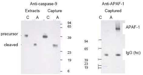
      Figure 2: Caspase-9 complexed with APAF-1 is captured in the Active Caspase-9 Assay Kit

      Jurkat cell extracts were activated for 60 min at 30°C with cytochrome c and dATP (A) or with buffer only (C). Extracts were then incubated in plate wells coated with Caspase-9 Capture Antibody for 2 h. Wells were washed and captured proteins were solubilized in SDS sample buffer. Immunoblots of extracts and captured proteins were performed using an anti-caspase-9 antibody or an anti-APAF-1 (Cat. No. AP1038) antibody.

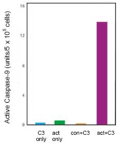
      Figure 3: Activated Extract and Precursor Caspase-3 Are Required for DEVD-AFC Cleavage

      Caspase-9 in activated and control Jurkat cell extracts (5 x 10⁵ cells) were captured in wells coated with Caspase-9 Capture Antibody. Wells were washed and Precursor Caspase-3 was added. After a 60 min incubation, cleavage of DEVD-AFC by active caspase-3 was measured using a fluorescent plate reader. Precursor Caspase-3 only (C3 only), activated extracts only (act only), a control extract with Precursor Caspase-3 (con+C3), and activated extract with Precursor Caspase-3 (act+C3) were employed in this assay. A unit of caspase-9 activity is defined as the amount of active caspase-9 required to activate sufficient caspase-3 to cleave DEVD-AFC at a rate of 1 RFU/min.

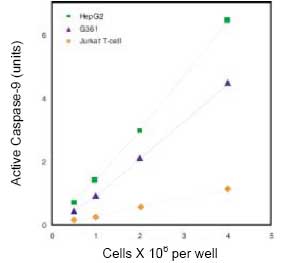
      Figure 4: Active Caspase-9 is Proportional to the Amount of Activated Cell Extracts Assaye

      Human HepG2 hepatocytes, G361 melanoma cells, and Jurkat T cells were solubilized in Caspase-9 Lysis Buffer 1 at a concentration of 2 x 10⁸ cells/ml. Caspase-9 was activated by incubating the indicated amount of cell lysates with cytochrome c and dATP. Activated lysates were analyzed using the Active Caspase-9 Assay Kit.

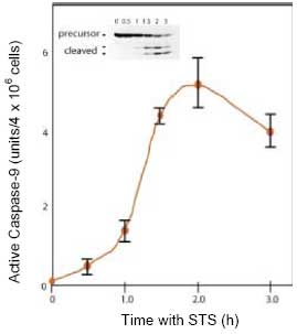
      Figure 5: The Active Caspase-9 Assay Kit Detects Caspase-9 Activated in Cultured Cells

      Jurkat cells were incubated with 1 µM staurosporine (STS) for the indicated times. Cells were rinsed with PBS and solubilized at 2 x 10⁸ cells/ml in Caspase-9 Lysate Buffer 2. Lysates were analyzed using the Active Caspase-9 assay Kit and by immunoblotting using a caspase-9 specific antibody. The caspase-9 activity detected in this assay correlates well with the amount of caspase-9 cleavage observed by immunoblotting.

      Registered TrademarksCalbiochem® is a registered trademark of EMD Chemicals, Inc. Tween® is a registered trademark of ICI Americas, Inc. Interactive Pathways™ is a trademark of EMD Chemicals, Inc.

      Related Products & Applications

      Isalterdis

      Catalogue Number Description  
      QIA72 Caspase-9 Activity Assay Kit, Fluorometric Show Pricing & Availability

      Categories

      Life Science Research > Protein Detection and Quantification