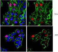
Single-Cell Profiling of Epigenetic Modifiers Identifies PRDM14 as an Inducer of Cell Fate in the Mammalian Embryo.
Burton, Adam, et al.
Cell Rep, (2013)
2013
Show Abstract
Cell plasticity or potency is necessary for the formation of multiple cell types. The mechanisms underlying this plasticity are largely unknown. Preimplantation mouse embryos undergo drastic changes in cellular potency, starting with the totipotent zygote through to the formation of the pluripotent inner cell mass (ICM) and differentiated trophectoderm in the blastocyst. Here, we set out to identify and functionally characterize chromatin modifiers that define the transitions of potency and cell fate in the mouse embryo. Using a quantitative microfluidics approach in single cells, we show that developmental transitions are marked by distinctive combinatorial profiles of epigenetic modifiers. Pluripotent cells of the ICM are distinct from their differentiated trophectoderm counterparts. We show that PRDM14 is heterogeneously expressed in 4-cell-stage embryos. Forced expression of PRDM14 at the 2-cell stage leads to increased H3R26me2 and can induce a pluripotent ICM fate. Our results shed light on the epigenetic networks that govern cellular potency and identity in vivo. VIDEO ABSTRACT: | Immunocytochemistry | | 24183668
 |
Ablation of PRMT6 reveals a role as a negative transcriptional regulator of the p53 tumor suppressor.
Neault, M; Mallette, FA; Vogel, G; Michaud-Levesque, J; Richard, S
Nucleic acids research
40
9513-21
2012
Show Abstract
Arginine methylation of histones is a well-known regulator of gene expression. Protein arginine methyltransferase 6 (PRMT6) has been shown to function as a transcriptional repressor by methylating the histone H3 arginine 2 [H3R2(me2a)] repressive mark; however, few targets are known. To define the physiological role of PRMT6 and to identify its targets, we generated PRMT6(-/-) mouse embryo fibroblasts (MEFs). We observed that early passage PRMT6(-/-) MEFs had growth defects and exhibited the hallmarks of cellular senescence. PRMT6(-/-) MEFs displayed high transcriptional levels of p53 and its targets, p21 and PML. Generation of PRMT6(-/-); p53(-/-) MEFs prevented the premature senescence, suggesting that the induction of senescence is p53-dependent. Using chromatin immunoprecipitation assays, we observed an enrichment of PRMT6 and H3R2(me2a) within the upstream region of Trp53. The PRMT6 association and the H3R2(me2a) mark were lost in PRMT6(-/-) MEFs and an increase in the H3K4(me3) activator mark was observed. Our findings define a new regulator of p53 transcriptional regulation and define a role for PRMT6 and arginine methylation in cellular senescence. | Western Blotting | | 22904064
 |
Analysis of histones in Xenopus laevis. I. A distinct index of enriched variants and modifications exists in each cell type and is remodeled during developmental transitions.
Shechter, D; Nicklay, JJ; Chitta, RK; Shabanowitz, J; Hunt, DF; Allis, CD
The Journal of biological chemistry
284
1064-74
2009
Show Abstract
Histone proteins contain epigenetic information that is encoded both in the relative abundance of core histones and variants and particularly in the post-translational modification of these proteins. We determined the presence of such variants and covalent modifications in seven tissue types of the anuran Xenopus laevis, including oocyte, egg, sperm, early embryo equivalent (pronuclei incubated in egg extract), S3 neurula cells, A6 kidney cells, and erythrocytes. We first developed a new robust method for isolating the stored, predeposition histones from oocytes and eggs via chromatography on heparin-Sepharose, whereas we isolated chromatinized histones via conventional acid extraction. We identified two previously unknown H1 isoforms (H1fx and H1B.Sp) present on sperm chromatin. We immunoblotted this global collection of histones with many specific post-translational modification antibodies, including antibodies against methylated histone H3 on Lys(4), Lys(9), Lys(27), Lys(79), Arg(2), Arg(17), and Arg(26); methylated histone H4 on Lys(20); methylated H2A and H4 on Arg(3); acetylated H4 on Lys(5), Lys(8), Lys(12), and Lys(16) and H3 on Lys(9) and Lys(14); and phosphorylated H3 on Ser(10) and H2A/H4 on Ser(1). Furthermore, we subjected a subset of these histones to two-dimensional gel analysis and subsequent immunoblotting and mass spectrometry to determine the global remodeling of histone modifications that occurs as development proceeds. Overall, our observations suggest that each metazoan cell type may have a unique histone modification signature correlated with its differentiation status. | | | 18957438
 |
Redundant requirement for a pair of PROTEIN ARGININE METHYLTRANSFERASE4 homologs for the proper regulation of Arabidopsis flowering time.
Niu, L; Zhang, Y; Pei, Y; Liu, C; Cao, X
Plant physiology
148
490-503
2008
Show Abstract
CARM1/PRMT4 (for COACTIVATOR-ASSOCIATED ARGININE METHYLTRANSFERASE1/PROTEIN ARGININE METHYLTRANSFERASE4) catalyzes asymmetric dimethylation on arginine (Arg), and its functions in gene regulation is understood only in animal systems. Here, we describe AtPRMT4a and AtPRMT4b as a pair of Arabidopsis (Arabidopsis thaliana) homologs of mammalian CARM1/PRMT4. Recombinant AtPRMT4a and AtPRMT4b could asymmetrically dimethylate histone H3 at Arg-2, Arg-17, Arg-26, and myelin basic protein in vitro. Both AtPRMT4a and AtPRMT4b exhibited nuclear as well as cytoplasmic distribution and were expressed ubiquitously in all tissues throughout development. Glutathione S-transferase pull-down assays revealed that AtPRMT4a and AtPRMT4b could form homodimers and heterodimers in vitro, and formation of the heterodimer was further confirmed by bimolecular fluorescence complementation. Simultaneous lesions in AtPRMT4a and AtPRMT4b genes led to delayed flowering, whereas single mutations in either AtPRMT4a or AtPRMT4b did not cause major developmental defects, indicating the redundancy of AtPRMT4a and AtPRMT4b. Genetic analysis also indicated that atprmt4a atprmt4b double mutants phenocopied autonomous pathway mutants. Finally, we found that asymmetric methylation at Arg-17 of histone H3 was greatly reduced in atprmt4a atprmt4b double mutants. Taken together, our results demonstrate that AtPRMT4a and AtPRMT4b are required for proper regulation of flowering time mainly through the FLOWERING LOCUS C-dependent pathway. Full Text Article | Western Blotting | | 18660432
 |
Coactivator-associated arginine methyltransferase 1 (CARM1) is a positive regulator of the Cyclin E1 gene.
El Messaoudi, S; Fabbrizio, E; Rodriguez, C; Chuchana, P; Fauquier, L; Cheng, D; Theillet, C; Vandel, L; Bedford, MT; Sardet, C
Proceedings of the National Academy of Sciences of the United States of America
103
13351-6
2006
Show Abstract
The Cyclin E1 gene (CCNE1) is an ideal model to explore the mechanisms that control the transcription of cell cycle-regulated genes whose expression rises transiently before entry into S phase. E2F-dependent regulation of the CCNE1 promoter was shown to correlate with changes in the level of H3-K9 acetylation/methylation of nucleosomal histones positioned at the transcriptional start site region. Here we show that, upon growth stimulation, the same region is subject to variations of H3-R17 and H3-R26 methylation that correlate with the recruitment of coactivator-associated arginine methyltransferase 1 (CARM1) onto the CCNE1 and DHFR promoters. Accordingly, CARM1-deficient cells lack these modifications and present lowered levels and altered kinetics of CCNE1 and DHFR mRNA expression. Consistently, reporter gene assays demonstrate that CARM1 functions as a transcriptional coactivator for their E2F1/DP1-stimulated expression. CARM1 recruitment at the CCNE1 gene requires activator E2Fs and ACTR, a member of the p160 coactivator family that is frequently overexpressed in human breast cancer. Finally, we show that grade-3 breast tumors present coelevated mRNA levels of ACTR and CARM1, along with their transcriptional target CCNE1. All together, our results indicate that CARM1 is an important regulator of the CCNE1 gene. | | | 16938873
 |
Chromatin of the Barr body: histone and non-histone proteins associated with or excluded from the inactive X chromosome.
Chadwick, BP; Willard, HF
Human molecular genetics
12
2167-78
2003
Show Abstract
The Barr body has long been recognized as the cytological manifestation of the inactive X chromosome (Xi) in interphase nuclei. Despite being known for over 50 years, relatively few components of the Barr body have been identified. In this study, we have screened over 30 histone variants, modified histones and non-histone proteins for their association with or exclusion from the Barr body. We demonstrate that, similar to the histone variant macroH2A, heterochromatin protein-1 (HP1), histone H1 and the high mobility group protein HMG-I/Y are elevated at the territory of the Xi in interphase in human cell lines, but only when the Xi chromatin is heteropycnotic, implicating each as a component of the Barr body. Surprisingly, however, virtually all other candidate proteins involved in establishing heterochromatin and gene silencing are notably absent from the Barr body despite being localized generally elsewhere throughout the nucleus, indicating that the Barr body represents a discrete subnuclear compartment that is not freely accessible to most chromatin proteins. A similar dichotomous pattern of association or exclusion describes the spatial relationship of a number of specific histone methylation patterns in relation to the Barr body. Notably, though, several methylated forms of histone H3 that are deficient in Xi chromatin generally are present at a region near the macrosatellite repeat DXZ4, as are the chromatin proteins CTCF and SAP30, indicating a distinctive chromatin state in this region of the Xi. Taken together, our data imply that the Xi adopts a distinct chromatin configuration in interphase nuclei and are consistent with a mechanism by which HP1, through histone H3 lysine-9 methylation, recognizes and assists in maintaining heterochromatin and gene silencing at the human Xi. | | Human | 12915472
 |


