Signaling by the Epstein-Barr virus LMP1 protein induces potent cytotoxic CD4+ and CD8+ T cell responses.
Choi, IK; Wang, Z; Ke, Q; Hong, M; Qian, Y; Zhao, X; Liu, Y; Kim, HJ; Ritz, J; Cantor, H; Rajewsky, K; Wucherpfennig, KW; Zhang, B
Proc Natl Acad Sci U S A
115
E686-E695
2018
Show Abstract
The B-lymphotropic Epstein-Barr virus (EBV), pandemic in humans, is rapidly controlled on initial infection by T cell surveillance; thereafter, the virus establishes a lifelong latent infection in the host. If surveillance fails, fatal lymphoproliferation and lymphomagenesis ensue. The initial T cell response consists of predominantly CD8+ cytotoxic T cells and a smaller expansion of CD4+ cells. A major approach to treating EBV-associated lymphomas is adoptive transfer of autologous or allogeneic T cells that are stimulated/expanded on EBV-transformed B cells. Strikingly, the clinical response correlates with the frequency of CD4 cells in the infused T cells. Although in vitro studies suggested that EBV-specific CD4 cells develop cytotoxicity, they have not been comprehensively characterized and the molecular mechanism underlying their formation remains unknown. Our recent work, using a transgenic approach in mice, has revealed a central role for the EBV signaling molecule LMP1 in immune surveillance and transformation of EBV-infected B cells. The mouse model offers a unique tool for uncovering basic features of EBV immunity. Here, we show that LMP1 expression in B cells induces potent cytotoxic CD4 and CD8 T cell responses, by enhancing antigen presentation and costimulation by CD70, OX40 ligand, and 4-1BB ligand. Our data further suggest that cytotoxic CD4 cells hold superior therapeutic value for LMP1 (EBV)-driven lymphomas. These findings provide insights into EBV immunity, demonstrating that LMP1 signaling alone is sufficient to induce a prominent cytotoxic CD4 response, and suggest strategies for immunotherapy in EBV-related and other cancers. | 29311309
 |
EBV latent membrane protein 1 abundance correlates with patient age but not with metastatic behavior in north African nasopharyngeal carcinomas.
Khabir, A; Karray, H; Rodriguez, S; Rosé, M; Daoud, J; Frikha, M; Boudawara, T; Middeldorp, J; Jlidi, R; Busson, P
Virol J
2
39
2005
Show Abstract
Undifferentiated nasopharyngeal carcinomas are rare in a majority of countries but they occur at a high incidence in South China and to a lesser extent in North Africa. They are constantly associated with the Epstein-Barr virus (EBV) regardless of patient geographic origin. In North Africa, the distribution of NPC cases according to patient age is bi-modal with a large group of patients being around 50 years old (80%) and a smaller group below 25 years old. We and others have previously shown that the juvenile form of NPC has distinct biological characteristics including a low amount of p53 and Bcl2 in the tumor tissue and a low level of anti-EBV IgG and IgA in the peripheral blood.To get more insight on potential oncogenic mechanisms specific of these two forms, LMP1 abundance was assessed in 82 NPC patients of both groups, using immuno-histochemistry and semi-quantitative evaluation of tissue staining. Serum levels of anti-EBV antibodies were simultaneously assessed. For LMP1 staining, we used the S12 antibody which has proven to be more sensitive than the common anti-LMP1 CS1-4 for analysis of tissue sections. In all NPC biopsies, at least a small fraction of cells was positively stained by S12. LMP1 abundance was strongly correlated to patient age, with higher amounts of the viral protein detected in specimens of the juvenile form. In contrast, LMP1 abundance was not correlated to the presence of lymph node or visceral metastases, nor to the risk of metastatic recurrence. It was also independent of the level of circulating anti-EBV antibodies.The high amount of LMP1 recorded in tumors from young patients confirms that the juvenile form of NPC has specific features regarding not only cellular but also viral gene expression. | 15842731
|
Preferential localization of the Epstein-Barr virus (EBV) oncoprotein LMP-1 to nuclei in human T cells: implications for its role in the development of EBV genome-positive T-cell lymphomas.
Xu, J; Ahmad, A; Menezes, J
J Virol
76
4080-6
2002
Show Abstract
The Epstein-Barr virus (EBV)-encoded latent membrane protein-1 (LMP-1) is thought to play a role in the EBV-induced B-cell transformation and immortalization. EBV has also been implicated in certain human T-cell lymphomas; however, the phenotypic effects of the expression of this oncoprotein in T cells are not known. To learn whether LMP-1 also induces phenotypic changes in T cells, we stably expressed it in human cell lines of T and B lineages and 25 LMP-1-expressing T-cell clones and 7 B-cell clones were examined. Our results show for the first time that, in sharp contrast to B cells, LMP-1 preferentially localizes to nuclei in T cells and does not induce the phenotypic changes in these cells that it induces in B cells, does not associate with TRAF proteins, and does not arrest the cell cycle in the G2/M phase. A computer-assisted analysis revealed that LMP-1 lacks the canonical nuclear localization signal. Our results suggest that this oncoprotein may not play the same role in the lymphomagenesis of T cells as it does in B cells. | 11907247
|
Presence of Epstein-Barr virus latency type III at the single cell level in post-transplantation lymphoproliferative disorders and AIDS related lymphomas.
Brink, AA; Dukers, DF; van den Brule, AJ; Oudejans, JJ; Middeldorp, JM; Meijer, CJ; Jiwa, M
J Clin Pathol
51
911-9
1998
Show Abstract
To investigate the expression pattern of Epstein-Barr virus (EBV) latent genes at the single cell level in post-transplantation lymphoproliferative disorders and acquired immunodefiency syndrome (AIDS) related lymphomas, in relation to cellular morphology.Nine post-transplantation lymphoproliferative disorders and three AIDS related lymphomas were subjected to immunohistochemistry using monoclonal antibodies specific for EBV nuclear antigen 1 (EBNA1) (2H4), EBNA2 (PE2 and the new rat anti-EBNA2 monoclonal antibodies 1E6, R3, and 3E9), and LMP1 (CS1-4 and S12). Double staining was performed combining R3 or 3E9 with S12.R3 and 3E9 anti-EBNA2 monoclonal antibodies were more sensitive than PE2, enabling the detection of more EBNA2 positive lymphoma cells. Both in post-transplantation lymphoproliferative disorders and AIDS related lymphomas, different expression patterns were detected at the single cell level. Smaller neoplastic cells were positive for EBNA2 but negative for LMP1. Larger and more blastic neoplastic cells, sometimes resembling Reed-Sternberg cells, were LMP1 positive but EBNA2 negative (EBV latency type II). Morphologically intermediate neoplastic cells coexpressing EBNA2 and LMP1 (EBV latency type III), were detected using R3 and 3E9, and formed a considerable part of the neoplastic population in four of nine post-transplantation lymphoproliferative disorders and two of three AIDS related lymphomas. All samples contained a subpopulation of small tumour cells positive exclusively for Epstein-Barr early RNA and EBNA1. The relation between cellular morphology and EBV expression patterns in this study was less pronounced in AIDS related lymphomas than in post-transplantation lymphoproliferative disorders, because the AIDS related lymphomas were less polymorphic than the post-transplantation lymphoproliferative disorders.In post-transplantation lymphoproliferative disorders and AIDS related lymphomas, EBV latency type III can be detected by immunohistochemistry in a subpopulation of tumour cells using sensitive monoclonal antibodies R3 and 3E9. Our data suggest that EBV infected tumour cells in these lymphomas undergo gradual changes in the expression of EBV latent genes, and that these changes are associated with changes in cellular morphology. | 9462239
|
Epstein-Barr virus gene expression in nasopharyngeal carcinoma.
Young, LS; Dawson, CW; Clark, D; Rupani, H; Busson, P; Tursz, T; Johnson, A; Rickinson, AB
J Gen Virol
70 ( Pt 5)
1051-66
1989
Show Abstract
Epstein-Barr virus (EBV), an agent with growth transforming potential for human B cells, is associated with certain B cell lymphomas in man and also with an epithelial tumour, undifferentiated nasopharyngeal carcinoma (NPC). Since B cell growth transformation is associated with the constitutive expression of a small number of EBV-coded latent proteins, the nuclear antigens EBNA 1, EBNA 2, EBNA 3 and EBNA-LP and the latent membrane protein (LMP), the present work sought to determine whether this same pattern of virus gene expression occurred in NPC. Tumour biopsies were taken from NPC patients from three areas of differing tumour incidence (Kenya, Algeria, Britain) and immediately snap-frozen, as were biopsies of non-EBV-related carcinomas for controls. Immunoblotting of PAGE-separated proteins with selected human sera identified 24 NPC biopsies clearly expressing EBNA 1. When the analysis was extended using selected human sera with antibodies against the other EBNAs, there was no detectable expression of EBNA 2, EBNA 3 or EBNA-LP in any of these 24 biopsies; their EBNA 2-negative status was confirmed using a monoclonal antibody (MAb) PE2 which was reactive in immunoblotting and in immunoprecipitation with EBNA 2A and EBNA 2B proteins. Similar experiments with two different LMP-specific MAbs, CS1 to 4 and S12, revealed heterogeneity between NPC biopsies; 9/24 biopsies were demonstrably LMP-positive, the degree of expression varying considerably between individual tumours in a manner which was not related to the level of EBNA 1 expression. None of the 24 NPC biopsies expressed detectable amounts of EBV lytic cycle antigens. A nude mouse-passaged NPC cell line, C15, likewise expressed EBNA 1 and LMP but none of the other EBV latent proteins nor lytic cycle antigens. This work identifies a novel type of EBV-cell interaction in NPC cells which is distinct from that seen in in vitro transformed B cell lines and from that seen to date in EBV-positive B cell lymphomas. | 2836550
|
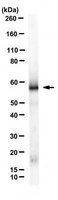
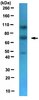
Epstein-Barr virus-encoded protein found in plasma membranes of transformed cells.
Mann, KP; Staunton, D; Thorley-Lawson, DA
J Virol
56
710-21
1986
Show Abstract
We have developed monoclonal antibodies to a 63,000-molecular-weight protein (p63) which is the product of the most abundant messenger RNA in Epstein-Barr virus-transformed cells and shown that the protein is associated specifically with plasma membranes. It was also found to be associated with the other membrane fractions and was found in all Epstein-Barr virus-transformed cells tested. In addition, p63 was present in virions, resulting in transient, early appearance in newly infected cells. Newly synthesized p63 was detected at the time cells underwent blast transformation (48 to 72 h postinfection). The possible role of this protein in transformation and as a target for cell-mediated cytotoxicity is discussed. | 2991591
|