Millipore Sigma Vibrant Logo
Attention: We have moved. Merck Millipore products are no longer available for purchase on MerckMillipore.com.Learn More

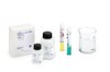
HCS17 c-fos Immunohistochemistry System

HCS17
  
Purchase on Sigma-Aldrich

Áttekintés

Replacement Information
Description
OverviewKit contains all required reagents for detecting Fos in paraffin-embedded tissues. The antibody will react with both v-Fos and c-Fos.
Catalogue NumberHCS17
Brand Family Calbiochem®
Materials Required but Not Delivered Coplin (or equivalent) staining jars Beakers Hanging slide racks Humidifying chamber Coverslips Light microscope Specimen: formalin-fixed, paraffin-embedded sections (4 to 8 microns thick) PBS: dissolve 8 g of NaCl (Cat. No. 567440), 200 mg of KCl (Cat. No. 529552), 1.44 g of Na2HPO4 (Cat. No. 567550 or 567547), and 240 mg KH2PO4 (Cat. No. 529565 or 529567) in 800 ml of distilled water; adjust the pH to 7.4 with HCl; add distilled water to 1 L. PBS-BSA: PBS with 1% (w/v) fatty acid free bovine serum albumin 1% Triton®X-100 detergent in PBS 30% H2O2 0.1% H2O2 (Cat. No. 386790) in distilled water, prepared fresh daily Solvents: 100% ethanol (Cat. No. 331542), 95% ethanol, xylene • Hematoxylin • Bluing reagent (optional) • Aqueous mounting medium (i.e. Permount®)
References
ReferencesDeTogni, P., et al. 1988. Mol. Cell Biol. 8, 2251. Rouscher III, F.J., et al. 1988. Science 240, 1010. Sassone-Corsi, P., et al. 1988. Cell 54, 553. Verma, I.M., and P. Sassone-Corsi. 1987. Cell 51, 513. Verma, I.M., and W.R. Graham. 1987. Adv. Cancer Res. 49, 29. Muller, R. 1986. Biochim. Biophys. Acta 823, 207. Verma, I. 1986. Trends Genet. 2, 93. Greenberg, M., and E. Ziff. 1984. Nature (London) 311, 433.
Product Information
Detection methodColorimetric
FormatMicroscopy
Kit containsPepsin, Primary Antibody c-Fos (Ab-2) (Cat. No. PC05), Negative Control Antibody, Blocking Serum, Biotinylated Secondary Antibody, Reagent A, Reagent B, DAB Tablets, Positive Control Slides, and a user protocol.
Positive controlIncluded in kit
Preservative≤0.1% sodium azide
Applications
Key Applications Paraffin Sections
Application CommentsA negative control antibody is also supplied and should be used at the same concentration. More intense staining may be obtained by preincubation with pepsin. Antibody should be titrated for optimal results in individual sample types. Materials Provided: • 100 µg c-fos antibody (Cat. No. PC05) • 100 µg normal rabbit IgG (Cat. No. NI01) for use as a negative control • goat serum (1 ml) for use as a blocker • biotinylated anti-rabbit IgG (1 ml) • Reagent A (avidin DH; 1 ml) • Reagent B (biotinylated horseradish peroxidase; 1 ml) • DAB Tablets (10 tablets, 10 mg DAB per tablet) • Pepsin (10 mg) • 2 slides of colon carcinoma tissue. Materials Required but not Provided: • Coplin (or equivalent) staining jars • Beakers • Hanging slide racks • Humidifying chamber • Coverslips • Light microscope • Specimen: formalin-fixed, paraffin-embedded sections (4 to 8 microns thick) • PBS: dissolve 8 g of NaCl (Cat. No. 567440), 200 mg of KCl (Cat. No. 529552), 1.44 g of Na2HPO4 (Cat. No. 567550 or 567547), and 240 mg KH2PO4 (Cat. No. 529565 or 529567) in 800 ml of distilled water; adjust the pH to 7.4 with HCl; add distilled water to 1 L. • PBS-BSA: PBS with 1% (w/v) fatty acid free bovine serum albumin • 1% Triton®X-100 detergent in PBS • 30% H2O2 • 0.1% H2O2 (Cat. No. 386790) in distilled water, prepared fresh daily • Solvents: 100% ethanol (Cat. No. 331542), 95% ethanol, xylene • Hematoxylin • Bluing reagent (optional) • Aqueous mounting medium (i.e. Permount®) Preparation of Paraffin-Eembedded Tissue Sections Deparaffinization of Formalin-fixed, Paraffin-embedded Tissue Sections 1. Deparaffinize in xylene using 3 changes, 5 min each. 2. Hydrate with 100% ethanol using 2 changes, 5 min each. 3. Hydrate with 95% ethanol using 2 changes, 5 min each. 4. Rinse in distilled water. 5. Incubate for 30 min in 0.1% H2O2 to quench any endogenous peroxidase activity. 6. Rinse in distilled water. 7. Rinse in PBS using 3 changes, 5 min each. 8. (Optional) Follow procedure for pretreatment with Pepsin protease (see below). Enzyme Pretreatment of Paraffin-Embedded Tissue Sections (Optional) 1. Incubate sections for 10 to 20 min in 0.1% Pepsin (Cat. No. 516360) in 0.01 N HCl, pH 2.3 at room temperature. 2. Terminate digestion by repeated washing in distilled water. Immunohistochemical Staining Carry out incubations at room temperature or at 4°C in a humidified chamber. Use suction to remove reagents after each step, but avoid drying of specimens between steps. Use sufficient reagents to cover the specimen (usually 20 to 50 µl). 1. Add one (1) drop of goat serum to 1 ml of PBS and mix. 2. Incubate specimens with diluted blocking serum in PBS for 20 min. EXPLANATION: Suppresses non-specific binding of immunoglobulin. Insufficient blocking (i.e. too short of an incubation time or too low of a serum concentration in the blocking solution) will lead to increased backgrounds. 3. Rinse with three changes of PBS, 5 min each rinse. 4. Incubate with Anti-c-fos antibody (Cat. No. PC05) or negative control antibody for either 60 min at room temperature or overnight at 4°C. Use negative control antibody at the same concentration as the primary antibody. For optimal results, incubation should be done overnight at 4°C in a humidified chamber. 5. Rinse with three changes of PBS, 5 min each rinse. Perform the next step during these incubations. 6. Prepare biotinylated secondary antibody by adding one (1) drop of stock to 1 ml PBS, then mix. Incubate with biotinylated second step antibody for 30 min at room temperature. Perform the next step during this incubation. 7. Prepare ABC Reagent by adding one (1) drop of Reagent A to 1 ml of buffer. Then add one (1) drop of Reagent B, mix immediately and let stand for 30 min before use at room temperature. EXPLANATION: The avidin DH:biotinylated horseradish peroxidase H complex consists of many biotinylated horseradish peroxidase molecules cross-linked by avidin to form a large macromolecular three dimensional array. At least one biotin binding site remains available to the biotinylated second step detector antibody. Incubation for 30 min ensures that an optimal complex is formed. Rinse with three changes of PBS, 5 min each rinse. 8. Incubate with ABC Reagent for 30 min. EXPLANATION: The pre-formed avidin-biotin(HRP) complex binds to the biotin on the secondary antibody and a lattice is formed. This localizes the HRP to the areas where the primary antibody has specifically bound to its antigen. 9. Rinse with three changes of PBS, 5 min each rinse. Perform the next step during these incubations. 10. Prepare DAB solution. Dilute as per product instructions or dissolve 5 mg DAB in 100 ml PBS together with 0.3 ml of 0.1% H2O2. CAUTION: DAB is a suspected carcinogen; use appropriate precautions. 11. Rinse in 1% Triton®X-100 detergent in PBS for 30 sec. 12. Incubate in DAB solution for 1-3 min. Check under microscope for development of brown color. If further intensification of staining is required, return slide to DAB solution and incubate an additional 1-5 min. EXPLANATION: The horseradish peroxidase converts the DAB substrate into an insoluble dark brown precipitate that is deposited around the binding site of the primary antibody. The presence of DAB localizes the site of expression of the antigen within the tissue section. 13. Rinse in distilled water. 14. Counter stain in Hematoxylin solution for 1-3 min. Duration will depend on the strength of Hematoxylin solution and degree of counterstaining desired. EXPLANATION: Hematoxylin stains the nucleus of the cells a purple color. This provides contrast to the brown DAB staining and permits the cellular architecture to be seen. 15. (Optional) Place the slides in Bluing reagent for 1 min. EXPLANATION: Bluing reagent turns the Hematoxylin stain a blue color providing a stronger contrast with the brown DAB staining. 16. Rinse in distilled water. Incubate in 95% ethanol, two changes, 10 s each. Incubate in 100% ethanol, two changes, 10 s each. Incubate in xylene, 2 changes, 10 s each. EXPLANATION: Removes excess water and causes the tissue to shrink slightly, enhancing the intensity of the staining. 17. Mount coverslips using aqueous mounting medium. 18. Examine by light microscopy. Trouble Shooting 1. No "brown" staining • Were the primary and secondary antibodies added? • Concentration of primary antibody may be too low. Increase the concentration by a factor of 2-3. • Make sure the H2O2 was added to the DAB solution. • Was the enzyme pretreatment step performed? • Slides left too short a time in the DAB solution. • Is the tissue type known to express the antigen? • Pretreatment was too long and the epitope was destroyed. Reduce the incubation time. • Were the antibodies stored at the correct temperature? Antibodies sold in solution will be inactivated by freezing, while antibodies sold lyophilized are best stored frozen after they are reconstituted. • Is the tissue from a species recognized by the primary antibody? 2. "Brown" staining too light • Primary antibody concentration too low. Increase the concentration by a factor of 2-3. • Increase the incubation time with the primary antibody. • Slides left too short a time in the DAB solution. • Level of expression may be low for the specific tissue sample. • How old are the reagents/kit? Check expiration date(s). • Enhance staining by placing slides in 0.5% cupric sulfate in 0.15 M NaCl for 5 min. Rinse in deionized water, then counterstain with Hematoxylin. 3. Sections are too brown or entire section is brown • Primary antibody concentration too high. Decrease the concentration by a factor of 2-3. • Slides left in the DAB solution too long. Shorten the incubation time in DAB. • Tissue was not properly blocked. Increase the incubation time with the blocking serum and/or increase the concentration of blocking serum by a factor of 3. • Washing steps in between incubations with antibody were shortened or omitted altogether. • Preincubation with H2O2 blocking solution was not performed. • H2O2 is old. New reagent should be obtained. • Cross-reactivity between the primary and/or secondary antibody and proteins in the tissue may be occurring. Add 1% normal rabbit serum to the blocking solution or 0.01% Triton®X-100 detergent to the primary and secondary antibody solutions (caution: this may also reduce specific binding of some antibodies). • Red blood cells in tissue sections can leave iron deposits that stain brown. Perfuse tissue before fixation. • Endogenous biotin in tissue can bind ABC reagents. Block biotin (i.e. with a commercially available kit). 4. Debris found on the slides • DAB is old and beginning to precipitate out of solution. Filter DAB solution before using. • Tissue sections are of poor quality. New sections should be obtained. • Xylene is dirty and should be changed. 5. No counterstaining or counterstaining is too light • Increase time left in the Hematoxylin solution. • Use a "bluing" reagent to enhance the staining. 6. Counterstain is too dark (no contrast is seen between the counterstain and DAB) • Sections left in Hematoxylin solution too long. Reduce the incubation period. • Insufficient incubation with DAB. Increase the incubation time in DAB. • Try an alternate histological counterstain such as methyl green. • Place sections in 70% ethanol with 1% (1 N) HCl until stain lightens. 7. Staining pattern not what is expected (cytoplasmic as well as nuclear) • Tissue was over digested with the enzyme pretreatment step. Reduce the duration of the incubation or the concentration of enzyme used. • Sections were improperly blocked with the serum blocker. Increase incubation time for the blocking step or increase the concentration of blocker used. • Excess primary antibody was used. Decrease the concentration of primary antibody. • Cross-reactivity between the secondary antibody and proteins in the tissue may be occurring. Add 1% normal rabbit serum to the blocking solution or reduce the amount of secondary antibody by a factor of 3.
Biological Information
Assay time2 h
HostRabbit
Physicochemical Information
Dimensions
Materials Information
Toxicological Information
Safety Information according to GHS
Safety Information
Product Usage Statements
Storage and Shipping Information
Ship Code Blue Ice Only
Toxicity Multiple Toxicity Values, refer to MSDS
Storage +2°C to +8°C
Do not freeze Yes
Packaging Information
Transport Information
Supplemental Information
Kit containsPepsin, Primary Antibody c-Fos (Ab-2) (Cat. No. PC05), Negative Control Antibody, Blocking Serum, Biotinylated Secondary Antibody, Reagent A, Reagent B, DAB Tablets, Positive Control Slides, and a user protocol.
Specifications
Global Trade Item Number
Katalógusszám GTIN
HCS17 0

Documentation

References

Hivatkozások áttekintése
DeTogni, P., et al. 1988. Mol. Cell Biol. 8, 2251. Rouscher III, F.J., et al. 1988. Science 240, 1010. Sassone-Corsi, P., et al. 1988. Cell 54, 553. Verma, I.M., and P. Sassone-Corsi. 1987. Cell 51, 513. Verma, I.M., and W.R. Graham. 1987. Adv. Cancer Res. 49, 29. Muller, R. 1986. Biochim. Biophys. Acta 823, 207. Verma, I. 1986. Trends Genet. 2, 93. Greenberg, M., and E. Ziff. 1984. Nature (London) 311, 433.

Brochure

Title
Caspases and other Apoptosis Related Tools Brochure
Kit SourceBook - 2nd Edition EURO
User Protocol

Revision20-October-2008 RFH
FormatMicroscopy
Detection methodColorimetric
Specieshuman, mouse, rat
BackgroundThe proto-oncogene fos has been implicated in cell growth, differentiation and development. Proto-oncogene fos is induced by a large number of stimuli ranging from mitogens, differentiation-specific agents, pharmacological agents etc. Induction of the 62,000 dalton fos protein is rapid but transient. The fos protein is associated with a 39,000 dalton protein which has been identified as the protein product of the jun proto-oncogene (c-jun). The fos/jun protein complex binds specifically to a sequence element in DNA referred to as the AP-1 binding site.
Materials provided• 100 µg c-fos antibody (Cat. No. PC05-100UG) • 100 µg normal rabbit IgG (Cat. No. NI01-100UG) for use as a negative control • goat serum (Kit Component No. JA1120-1ML): 1 ml for use as a blocker • biotinylated anti-rabbit IgG (Kit Component No. JA1130-1ML): 1 ml • Reagent A (avidin DH;) (Kit Component No. JA1070-1ML): 1 ml • Reagent B (biotinylated horseradish peroxidase;) (Kit Component No. JA1080-1ML): 1 ml • DAB Tablets (Kit Component No. JA1415-10EA): 10 tablets, 10 mg DAB per tablet • Pepsin (Kit Component No. JA1411-10MG): 10 mg • Slides Tissue Sections colon carcinoma Tissue (Kit Component No. JA1369-2EA): 2 slides
Materials Required but not provided Coplin (or equivalent) staining jars Beakers Hanging slide racks Humidifying chamber Coverslips Light microscope Specimen: formalin-fixed, paraffin-embedded sections (4 to 8 microns thick) PBS: dissolve 8 g of NaCl (Cat. No. 567440), 200 mg of KCl (Cat. No. 529552), 1.44 g of Na2HPO4 (Cat. No. 567550 or 567547), and 240 mg KH2PO4 (Cat. No. 529565 or 529567) in 800 ml of distilled water; adjust the pH to 7.4 with HCl; add distilled water to 1 L. PBS-BSA: PBS with 1% (w/v) fatty acid free bovine serum albumin 1% Triton®X-100 detergent in PBS 30% H2O2 0.1% H2O2 (Cat. No. 386790) in distilled water, prepared fresh daily Solvents: 100% ethanol (Cat. No. 331542), 95% ethanol, xylene • Hematoxylin • Bluing reagent (optional) • Aqueous mounting medium (i.e. Permount®)
PreparationPreparation of Paraffin-Embedded Tissue Sections Deparaffinization of Formalin-fixed, Paraffin-embedded Tissue Sections 1. Deparaffinize in xylene using 3 changes, 5 min each. 2. Hydrate with 100% ethanol using 2 changes, 5 min each. 3. Hydrate with 95% ethanol using 2 changes, 5 min each. 4. Rinse in distilled water. 5. Incubate for 30 min in 0.1% H2O2 to quench any endogenous peroxidase activity. 6. Rinse in distilled water. 7. Rinse in PBS using 3 changes, 5 min each. 8. (Optional) Follow procedure for pretreatment with Pepsin protease (see below). Enzyme Pretreatment of Paraffin-Embedded Tissue Sections (Optional) 1. Incubate sections for 10 to 20 min in 0.1% Pepsin (Cat. No. 516360) in 0.01 N HCl, pH 2.3 at room temperature. 2. Terminate digestion by repeated washing in distilled water.
Detailed protocolImmunohistochemical Staining Carry out incubations at room temperature or at 4°C in a humidified chamber. Use suction to remove reagents after each step, but avoid drying of specimens between steps. Use sufficient reagents to cover the specimen (usually 20 to 50 µl). 1. Add one (1) drop of goat serum to 1 ml of PBS and mix. 2. Incubate specimens with diluted blocking serum in PBS for 20 min. EXPLANATION: Suppresses non-specific binding of immunoglobulin. Insufficient blocking (i.e. too short of an incubation time or too low of a serum concentration in the blocking solution) will lead to increased backgrounds. 3. Rinse with three changes of PBS, 5 min each rinse. 4. Incubate with Anti-c-fos antibody (Cat. No. PC05) or negative control antibody for either 60 min at room temperature or overnight at 4°C. Use negative control antibody at the same concentration as the primary antibody. For optimal results, incubation should be done overnight at 4°C in a humidified chamber. 5. Rinse with three changes of PBS, 5 min each rinse. Perform the next step during these incubations. 6. Prepare biotinylated secondary antibody by adding one (1) drop of stock to 1 ml PBS, then mix. Incubate with biotinylated second step antibody for 30 min at room temperature. Perform the next step during this incubation. 7. Prepare ABC Reagent by adding one (1) drop of Reagent A to 1 ml of buffer. Then add one (1) drop of Reagent B, mix immediately and let stand for 30 min before use at room temperature. EXPLANATION: The avidin DH:biotinylated horseradish peroxidase H complex consists of many biotinylated horseradish peroxidase molecules cross-linked by avidin to form a large macromolecular three dimensional array. At least one biotin binding site remains available to the biotinylated second step detector antibody. Incubation for 30 min ensures that an optimal complex is formed. Rinse with three changes of PBS, 5 min each rinse. 8. Incubate with ABC Reagent for 30 min. EXPLANATION: The pre-formed avidin-biotin(HRP) complex binds to the biotin on the secondary antibody and a lattice is formed. This localizes the HRP to the areas where the primary antibody has specifically bound to its antigen. 9. Rinse with three changes of PBS, 5 min each rinse. Perform the next step during these incubations. 10. Prepare DAB solution. Dilute as per product instructions or dissolve 5 mg DAB in 100 ml PBS together with 0.3 ml of 0.1% H2O2. CAUTION: DAB is a suspected carcinogen; use appropriate precautions. 11. Rinse in 1% Triton®X-100 detergent in PBS for 30 sec. 12. Incubate in DAB solution for 1-3 min. Check under microscope for development of brown color. If further intensification of staining is required, return slide to DAB solution and incubate an additional 1-5 min. EXPLANATION: The horseradish peroxidase converts the DAB substrate into an insoluble dark brown precipitate that is deposited around the binding site of the primary antibody. The presence of DAB localizes the site of expression of the antigen within the tissue section. 13. Rinse in distilled water. 14. Counter stain in Hematoxylin solution for 1-3 min. Duration will depend on the strength of Hematoxylin solution and degree of counterstaining desired. EXPLANATION: Hematoxylin stains the nucleus of the cells a purple color. This provides contrast to the brown DAB staining and permits the cellular architecture to be seen. 15. (Optional) Place the slides in Bluing reagent for 1 min. EXPLANATION: Bluing reagent turns the Hematoxylin stain a blue color providing a stronger contrast with the brown DAB staining. 16. Rinse in distilled water. Incubate in 95% ethanol, two changes, 10 s each. Incubate in 100% ethanol, two changes, 10 s each. Incubate in xylene, 2 changes, 10 s each. EXPLANATION: Removes excess water and causes the tissue to shrink slightly, enhancing the intensity of the staining. 17. Mount coverslips using aqueous mounting medium. 18. Examine by light microscopy.
Registered TrademarksCalbiochem® is a registered trademark of EMD Chemicals, Inc. Interactive Pathways™ is a trademark of EMD Chemicals, Inc.