Millipore Sigma Vibrant Logo
Attention: We have moved. Merck Millipore products are no longer available for purchase on MerckMillipore.com.Learn More

07-2177 Anti-phospho-PKD2 Antibody (Ser197/Ser200)

07-2177
100 µL  
Purchase on Sigma-Aldrich

Különleges ajánlatok

Áttekintés

Replacement Information

Különleges ajánlatok

Kulcsspecifikációk táblázata

Species ReactivityKey ApplicationsHostFormatAntibody Type
M, H, RWBRbAffinity PurifiedPolyclonal Antibody
Description
Catalogue Number07-2177
DescriptionAnti-phospho-PKD2 Antibody (Ser197/Ser200)
Alternate Names
  • Autosomal dominant polycystic kidney disease type II protein
  • Polycystic kidney disease 2 protein
  • Polycystwin
  • R48321
Background InformationAs members of the calcium-/calmodulin-depedent protein kinase superfamily, there are three known Protein Kinase D (PKD) isoforms, PKD1, PKD2, and PKD3. These serine/threonine kinases are involved in protein-protein and protein-carbohydrate adhesion and are also thought to be ion channel regulators. They are prominent downstream targets of protein kinase C and phospholipase D in various biological systems including mouse. PKDs are activated by various stimuli, including phorbol esters, G-protein-coupled receptors, reactive oxygen species, and diacylglycerol. PKDs regulate protein transport in the trans-Golgi network and are thought to play a role in muscle differentiation, cell motility, migration, and invasion. These isoforms are known to interact with each other. PKD2 is shown to be activated by phorbol esters, both in vivo and in vitro, and also by gastrin via the cholescystokinin/CCK(B) receptor in stably-transfected AGS-B cells. Furthermore, PKD2 is found to be active during the early phase of differentation of C2C12 cells into skeletal muscle cells. It is also reported to enhance TCR-induced NFAT activity in Jurkat T cells.
References
Product Information
FormatAffinity Purified
Control
  • MEF cell lysates untreated and PBDu-treated
PresentationPurified rabbit polyclonal in buffer containing 0.1 M Tris-Glycine (pH 7.4), 150 mM NaCl with 0.05% sodium azide.
Applications
Application
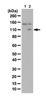
This phospho-PKD2 antibody is validated for use in WB for the detection of the phospho-PKD2 protein.
Key Applications
  • Western Blotting
Application NotesEvaluated by Western Blot in MEF cell lysates untreated and treated with PDBu, Rapamycin, and GO6983.

Western Blot Analysis: A 1:2,000 dilution of this antibody detected PKD2 in 10 µg of MEF cell lysates untreated and treated with PDBu, Rapamycin, and GO6983.
Biological Information
ImmunogenKLH-conjugated linear peptide corresponding to human PKD2 phosphorylated at Ser197/Ser200.
EpitopePhosphorylated Ser197/Ser200
ConcentrationPlease refer to the Certificate of Analysis for the lot-specific concentration.
HostRabbit
SpecificityThis antibody recognizes PKD2 phosphorylated at Ser197/Ser200.
Species Reactivity
  • Mouse
  • Human
  • Rat
Species Reactivity NoteDemonstrated to react with Mouse.
Predicted to react with Human and Rat based on 100% sequence homology.
Antibody TypePolyclonal Antibody
Entrez Gene Number
Entrez Gene SummaryThis gene encodes a member of the polycystin protein family. The encoded protein is a multi-pass membrane protein that functions as a calcium permeable cation channel, and is involved in calcium transport and calcium signaling in renal epithelial cells. This protein interacts with polycystin 1, and they may be partners in a common signaling cascade involved in tubular morphogenesis. Mutations in this gene are associated with autosomal dominant polycystic kidney disease type 2.
Gene Symbol
  • PC2
  • PKD4
  • Pc-2
  • APKD2
  • TRPP2
  • MGC138466
  • MGC138468
Modifications
  • Phosphorylation
  • Phosphorylation
Purification MethodAffinity Purfied
UniProt Number
UniProt SummaryFUNCTION: Functions as a calcium permeable cation channel. PKD1 and PKD2 may function through a common signaling pathway that is necessary for normal tubulogenesis.

SUBUNIT STRUCTURE: Forms homooligomers. Interacts with PKD1. PKD1 requires the presence of PKD2 for stable expression. Interacts with CD2AP. Interacts with HAX1.

SUBCELLULAR LOCATION: Multi-pass membrane protein Potential. Endoplasmic reticulum.
Molecular Weight~110 kDa observed. An uncharacterized band appears at ~150 kDa in some lysates.
Physicochemical Information
Dimensions
Materials Information
Toxicological Information
Safety Information according to GHS
Safety Information
Product Usage Statements
Quality AssuranceEvaluated by Western Blot in MEF cell lysates untreated and PBDu-treated.

Western Blot Analysis: A 1:2,000 dilution of this antibody detected PKD2 in 10 µg of MEF cell lysates untreated and PBDu-treated.
Usage Statement
  • Unless otherwise stated in our catalog or other company documentation accompanying the product(s), our products are intended for research use only and are not to be used for any other purpose, which includes but is not limited to, unauthorized commercial uses, in vitro diagnostic uses, ex vivo or in vivo therapeutic uses or any type of consumption or application to humans or animals.
Storage and Shipping Information
Storage ConditionsStable for 1 year at 2-8°C from date of receipt.
Packaging Information
Material Size100 µL
Transport Information
Supplemental Information
Specifications
Global Trade Item Number
Katalógusszám GTIN
07-2177 04053252420405