Millipore Sigma Vibrant Logo
Attention: We have moved. Merck Millipore products are no longer available for purchase on MerckMillipore.com.Learn More

MAB8151-I-100UL Anti-West Nile Virus/Kunjin Antibody, Envelope Antibody, clone 3.91D

MAB8151-I-100UL
100 µL  
Purchase on Sigma-Aldrich

Áttekintés

Replacement Information
Description
Catalogue NumberMAB8151-I-100UL
DescriptionAnti-West Nile Virus/Kunjin Antibody, Envelope Antibody, clone 3.91D
Alternate Names
  • Envelope protein
  • WNV Envelope Protein
Background InformationGenome polyprotein (UniProt: P14335) is encoded by the POLY gene (Gene ID: 912267) in West Nile virus. West Nile virus (WNV) is a mosquito-borne positive-sense RNA virus of the Flavivirus genus within the Flaviviridae family. WNV genome encodes for three structural domains known as C, prM, and E and seven non-structural proteins (NS1, NS2A, NS2B, NS3, NS4A, NS4B, and NS5). These are translated as a single polypeptide, which is subsequently cleaved by viral and cellular proteases. The envelope glycoprotein (E) undergoes a conformational rearrangement triggered by low pH that results in a class II fusion event required for viral entry. The E protein (aa 291-791) contains a three-domain structure where domain I is an eight-stranded β-barrel in the center portion of the protein (aa 1-51, 134-195, and 284-297). The DII domain consists of 170 residues inserted into DI (aa 52-133 and 196-283). At the distal end of DII domain is a fusion loop (aa 98-110) and a conserved glycine-rich, hydrophobic sequence, which is important for flaviviral fusion. The third domain, DIII, is an Ig-like domain with seven β-strand and has been implicated in receptor binding. The E protein also has a single N-linked glycosylation site at Asn 154, located within the ectodomain of prM domain and is cleaved by cellular enzymes during viral particle maturation. Clone E60 specifically binds to the DII fusion loop and displays limited neutralizing activity and even at higher doses neutralization activity is shown to be between 26 and 68%. (Ref.: Martín-Acebes MA, et al. (2012). World J Virol. 1(2); 51-70).
References
Product Information
FormatPurified
PresentationPurified mouse monoclonal antibody IgG3 in buffer containing 0.1 M Tris-Glycine (pH 7.4), 150 mM NaCl with 0.05% sodium azide.
Quality LevelMQ200
Applications
ApplicationAnti-West Nile Virus/Kunjin, Envelope, clone 3.91D, Cat. No. MAB8151-I, is a mouse monoclonal antibody that detects envelope protein in West Nile virus and is tested for use in ELISA, Immunofluorescence, and Western Blotting.
Key Applications
  • Western Blotting
  • Immunofluorescence
  • ELISA
Application NotesWestern Blotting Analysis: A representative lot detected West Nile Virus/Kunjin, Envelope protein in Western Blotting applications (Maeda, A., et al. (2009). Virus Res.;144(1-2):35-43; Saiyasombat, R., et al. (2014). Virol J.;11:150; Blitvich, B.J., et al. (2016). Am J Trop Med Hyg.;95(5):1185-1191).

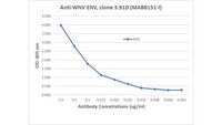
ELISA Analysis: Various dilutions of this antibody detected recombinant West Nile Virus/Kunjin, Envelope protein.

Immunofluorescence Analysis: A representative lot detected West Nile Virus/Kunjin, Envelope protein in Immunofluorescence applications (Maeda, A., et al. (2009). Virus Res.;144(1-2):35-43; Osorio, J.E., et al. (2012). Am J Trop Med Hyg.;87(3):565-72).

Note: Actual optimal working dilutions must be determined by end user as specimens, and experimental conditions may vary with the end user
Biological Information
ImmunogenWest Nile Virus (WNV; subtype KUNV) isolate OR393.
EpitopeN-terminal half
Clone3.91D
Concentration0.5 mg/mL. Please refer to guidance on suggested starting dilutions and/or titers per application and sample type.
HostMouse
SpecificityClone 3.91D is a mouse monoclonal antibody that detects West Nile virus envelope protein. It targets an epitope within the N-terminal half of polyprotein.
IsotypeIgG3
Species Reactivity
  • Virus
Species Reactivity NoteWest Nile virus.
Antibody TypeMonoclonal Antibody
Entrez Gene Number
Gene Symbol
  • E
Purification MethodProtein G purified
UniProt Number
Molecular Weight~50 kDa observed; 31.89 kDa calculated. Uncharacterized bands may be observed in some lysate(s).
Physicochemical Information
Dimensions
Materials Information
Toxicological Information
Safety Information according to GHS
Safety Information
Product Usage Statements
Quality AssuranceEvaluated by Western Blotting with recombinant West Nile Virus envelope protein.

Western Blotting Analysis (WB): A 1:500 dilution of this antibody detected recombinant West Nile Virus envelope protein.
Usage Statement
  • Unless otherwise stated in our catalog or other company documentation accompanying the product(s), our products are intended for research use only and are not to be used for any other purpose, which includes but is not limited to, unauthorized commercial uses, in vitro diagnostic uses, ex vivo or in vivo therapeutic uses or any type of consumption or application to humans or animals.
Storage and Shipping Information
Storage ConditionsRecommend storage at +2°C to +8°C. For long term storage antibodies can be kept at -20°C. Avoid repeated freeze-thaws.
Packaging Information
Material Size100 µL
Transport Information
Supplemental Information
Specifications
Global Trade Item Number
Katalógusszám GTIN
MAB8151-I-100UL 04065266791518