Bacterial lipoprotein TLR2 agonists broadly modulate endothelial function and coagulation pathways in vitro and in vivo.
Shin, HS; Xu, F; Bagchi, A; Herrup, E; Prakash, A; Valentine, C; Kulkarni, H; Wilhelmsen, K; Warren, S; Hellman, J
Journal of immunology (Baltimore, Md. : 1950)
186
1119-30
2010
Kivonat megmutatása
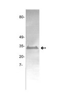
TLR2 activation induces cellular and organ inflammation and affects lung function. Because deranged endothelial function and coagulation pathways contribute to sepsis-induced organ failure, we studied the effects of bacterial lipoprotein TLR2 agonists, including peptidoglycan-associated lipoprotein, Pam3Cys, and murein lipoprotein, on endothelial function and coagulation pathways in vitro and in vivo. TLR2 agonist treatment induced diverse human endothelial cells to produce IL-6 and IL-8 and to express E-selectin on their surface, including HUVEC, human lung microvascular endothelial cells, and human coronary artery endothelial cells. Treatment of HUVEC with TLR2 agonists caused increased monolayer permeability and had multiple coagulation effects, including increased production of plasminogen activator inhibitor-1 (PAI-1) and tissue factor, as well as decreased production of tissue plasminogen activator and tissue factor pathway inhibitor. TLR2 agonist treatment also increased HUVEC expression of TLR2 itself. Peptidoglycan-associated lipoprotein induced IL-6 production by endothelial cells from wild-type mice but not from TLR2 knockout mice, indicating TLR2 specificity. Mice were challenged with TLR2 agonists, and lungs and plasmas were assessed for markers of leukocyte trafficking and coagulopathy. Wild-type mice, but not TLR2 mice, that were challenged i.v. with TLR2 agonists had increased lung levels of myeloperoxidase and mRNAs for E-selectin, P-selectin, and MCP-1, and they had increased plasma PAI-1 and E-selectin levels. Intratracheally administered TLR2 agonist caused increased lung fibrin levels. These studies show that TLR2 activation by bacterial lipoproteins broadly affects endothelial function and coagulation pathways, suggesting that TLR2 activation contributes in multiple ways to endothelial activation, coagulopathy, and vascular leakage in sepsis. | Western Blotting | 21169547
 |
Vascular biology-the role of tissue factor.
Hathcock, James
Semin. Hematol., 41: 30-4 (2004)
2004
Kivonat megmutatása
It is well established that tissue factor (TF) is abundantly present in various extravascular tissues, in the adventitia of blood vessels, and in atheroma. Thus, in the event of plaque rupture or damage to the blood vessel wall, TF is readily exposed to flowing blood, allowing it to form a complex with circulating factor VIIa (FVIIa) in order to activate factor X (FX) both directly, and indirectly via factor IX (FIX). Platelets quickly adhere to the injured site, facilitating localized thrombin formation and subsequent fibrin production. With each new layer of platelets and fibrin that adheres to the injured surface, the exposed TF on the vessel wall, along with the localized circulating factors IX (FIXa) and X (FXa) that it generates, becomes increasingly isolated from the events near the surface of the growing thrombus. The physical blanketing of an injured surface by platelets and fibrin in addition to the release of platelet tissue factor pathway inhibitor (TFPI), prevents FIXa and FXa from diffusing more than a few tens of microns away from the vessel wall, far short of the 3 mm thickness needed for occlusive thrombosis. Thus an alternative FXa-generating mechanism must be involved that allows for the formation of prothrombinase activity far away from the vessel wall near the front of a growing thrombus. | | 14872418
 |
Targeting tissue factor as an antithrombotic strategy.
Golino, Paolo and Cimmino, Giovanni
Seminars in vascular medicine, 3: 205-14 (2003)
2003
Kivonat megmutatása
It is generally accepted that the initial event in coagulation and intravascular thrombus formation is the exposure of cell-surface protein, such as tissue factor (TF). TF is exposed to the flowing blood as a consequence of vascular injury induced, for instance, by PTCA, or by spontaneous rupture of an atherosclerotic plaque. Expression of TF may also be induced in monocytes and endothelial cells in several conditions such as sepsis and cancer, causing a more generalized activation of clotting. In addition to its essential role in hemostasis, TF may be also implicated in several pathophysiological processes, such as intracellular signaling, cell proliferation, and inflammation. For all these reasons, TF has been the subject of intense research focus. Many experimental studies have demonstrated that inhibition of TF:factor VIIa procoagulant activity is a powerful inhibitor of in vivo thrombosis and that this approach usually results in a less-pronounced bleeding tendency compared with other "more classical" antithrombotic interventions. Alternative approaches may be represented by antibodies directed against TF, by transfection of the arterial wall with natural inhibitors of the TF:factor VIIa complex, such as the TF pathway inhibitor, or with catalytic RNA (ribozyme), which could inhibit the expression of the TF protein by the disruption of cellular TF mRNA. All these approaches seem particularly attractive because they may result in complete inhibition of local thrombosis without incurring potentially harmful systemic effects. Further studies are warranted to determine the efficacy and safety of such approaches in patients. | | 15199484
 |


