RNA-binding proteins TIA-1 and TIAR link the phosphorylation of eIF-2 alpha to the assembly of mammalian stress granules.
Kedersha, NL; Gupta, M; Li, W; Miller, I; Anderson, P
J Cell Biol
147
1431-42
1998
Kivonat megmutatása
In response to environmental stress, the related RNA-binding proteins TIA-1 and TIAR colocalize with poly(A)(+) RNA at cytoplasmic foci that resemble the stress granules (SGs) that harbor untranslated mRNAs in heat shocked plant cells (Nover et al. 1989; Nover et al. 1983; Scharf et al. 1998). The accumulation of untranslated mRNA at SGs is reversible in cells that recover from a sublethal stress, but irreversible in cells subjected to a lethal stress. We have found that the assembly of TIA-1/R(+) SGs is initiated by the phosphorylation of eIF-2alpha. A phosphomimetic eIF-2alpha mutant (S51D) induces the assembly of SGs, whereas a nonphosphorylatable eIF-2alpha mutant (S51A) prevents the assembly of SGs. The ability of a TIA-1 mutant lacking its RNA-binding domains to function as a transdominant inhibitor of SG formation suggests that this RNA-binding protein acts downstream of the phosphorylation of eIF-2alpha to promote the sequestration of untranslated mRNAs at SGs. The assembly and disassembly of SGs could regulate the duration of stress- induced translational arrest in cells recovering from environmental stress. | 10613902
 |
RNA-binding protein TIAR is essential for primordial germ cell development.
Beck, AR; Miller, IJ; Anderson, P; Streuli, M
Proc Natl Acad Sci U S A
95
2331-6
1998
Kivonat megmutatása
Primordial germ cells (PGCs) give rise to both eggs and sperm via complex maturational processes that require both cell migration and proliferation. However, little is known about the genes controlling gamete formation during the early stages of PGC development. Although several mutations are known to severely reduce the number of PGCs reaching and populating the genital ridges, the molecular identity of only two of these genes is known: the c-kit receptor protein tyrosine kinase and the c-kit ligand (the steel factor). Herein, we report that mutant mice lacking TIAR, an RNA recognition motif/ribonucleoprotein-type RNA-binding protein highly expressed in PGCs, fail to develop spermatogonia or oogonia. This developmental defect is a consequence of reduced survival of PGCs that migrate to the genital ridge around embryonic day 11.5 (E11.5). The numbers of PGCs populating the genital ridge in TIAR-deficient embryos are severely reduced compared to wild-type embryos by E11.5 and in the mutants PGCs are completely absent at E13.5. Furthermore, TIAR-deficient embryonic stem cells do not proliferate in the absence of exogenous leukemia inhibitory factor in an in vitro methylcellulose culture assay, supporting a role for TIAR in regulating cell proliferation. Because the development of PGCs relies on the action of several growth factors, these results are consistent with a role for TIAR in the expression of a survival factor or survival factor receptor that is essential for PGC development. TIAR-deficient mice thus provide a model system to study molecular mechanisms of PGC development and possibly the basis for some forms of idiopathic infertility. | 9482885
|
Structure, tissue distribution and genomic organization of the murine RRM-type RNA binding proteins TIA-1 and TIAR.
Beck, AR; Medley, QG; O'Brien, S; Anderson, P; Streuli, M
Nucleic Acids Res
24
3829-35
1996
Kivonat megmutatása
TIA-1 and TIAR are RNA binding proteins of the RNA recognition motif (RRM)/ribonucleoprotein (RNP) family that have been implicated as effectors of apoptotic cell death. We report the structures of murine TIA-1 and TIAR (mTIA-1 and mTIAR) deduced from cDNA cloning, the mRNA and protein tissue distribution of mTIA-1 and mTIAR, and the exon-intron structures of the mTIA-1 and mTIAR genes. Both mTIA-1 and mTIAR are comprised of three approximately 100 amino acid N-terminal RRM domains and a approximately 90 amino acid C-terminal auxiliary domain. This subfamily of RRM proteins is evolutionarily well conserved; mTIA-1 and mTIAR are 80% similar to each other, and 96 and 99% similar to hTIA-1 and hTIAR, respectively. The overall exon-intron structures of the mTIA-1 and mTIAR genes are also similar to each other, as well as to the human TIA-1 gene structure. While Northern blot analysis reveals that mTIA-1 and mTIAR mRNAs have a broad tissue distribution, mTIA-1 and mTIAR proteins are predominantly expressed in brain, testis and spleen. At least two isoforms of both mTIA-1 and mTIAR are generated by alternative splicing. Murine TIA-1 isoforms including or lacking the exon 5 encoded sequences are expressed at a ratio of approximately 1:1, whereas mTIAR isoforms including or lacking the 5'-end of exon 3 sequences are expressed in a approximately 1:6 ratio. Molecular characterization of murine TIA-1 and TIAR RNA binding proteins provides the basis for a genetic analysis of the functional roles of these proteins during mammalian development. | 8871565
|
The RNA-binding protein TIAR is translocated from the nucleus to the cytoplasm during Fas-mediated apoptotic cell death.
Taupin, JL; Tian, Q; Kedersha, N; Robertson, M; Anderson, P
Proc Natl Acad Sci U S A
92
1629-33
1994
Kivonat megmutatása
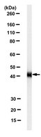
We have determined the structure, intracellular localization, and tissue distribution of TIAR, a TIA-1-related RNA-binding protein. Two related isoforms of TIAR, migrating at 42 and 50 kDa, are expressed in primate cells. Unlike TIA-1, which is found in the granules of cytotoxic lymphocytes, TIAR is concentrated in the nucleus of hematopoietic and nonhematopoietic cells. Because TIAR can trigger DNA fragmentation in permeabilized thymocytes, it is a candidate effector of apoptotic cell death. Consistent with this possibility, we have found that the expression and intracellular localization of TIAR change dramatically during Fas-mediated apoptosis. TIAR moves from the nucleus to the cytoplasm within 30 min of Fas ligation. Redistribution of TIAR precedes the onset of DNA fragmentation and is not a nonspecific consequence of nuclear disintegration. Cytoplasmic redistribution of TIAR is not observed during cellular activation triggered by mitogens such as concanavalin A or phytohemagglutinin. Our results suggest that cytoplasmic redistribution of TIAR may be a general feature of the apoptotic program. | 7533298
|