Millipore Sigma Vibrant Logo
Attention: We have moved. Merck Millipore products are no longer available for purchase on MerckMillipore.com.Learn More

AP1036 Anti-F₁F₀-α Mouse mAb (7H10BD4F9)

AP1036
  
Purchase on Sigma-Aldrich

Áttekintés

Replacement Information

Kulcsspecifikációk táblázata

Host
M
Description
Overview

This product has been discontinued.


Recognizes the F1F0-α subunit of Complex V in bovine heart mitochondria.
Catalogue NumberAP1036
Brand Family Calbiochem®
Application Data
Detection of bovine F1F0-α by immunoblotting. Sample: F1F0-α immunoprecipitated from heart mitochondria. Primary antibody: Anti-F1F0-α Mouse mAb (7H10BD4F9) (Cat. No. AP1036) (1 μg/ml). Detection: chemiluminescence.

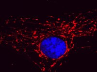
Detection of human F1F0-α by immunocytochemistry. Sample: 143B osteosarcoma cells. Primary antibody: Anti-F1F0-α Mouse mAb (7H10BD4F9) (Cat. No. AP1036) (5 μg/ml). Detection: fluorescence.
References
ReferencesMoser, T.L., et al. 2001. Proc. Natl. Acad. Sci. USA 98, 6656.
Osanai, T., et al. 2001. J. Clin. Invest. 108, 1023.
Moser, T.L., et al. 1999. Proc. Natl. Acad. Sci. USA 96, 2811.
Osanai, T., et al. 1998. J. Biol. Chem. 273, 31778.
Product Information
EC number3.6.1.14
FormLiquid
FormulationIn HEPES-buffered saline (HBS).
Positive controlHeart mitochondria
Preservative≤0.1% sodium azide
Applications
Key Applications Cell Inhibition Assays
Immunoblotting (Western Blotting)
Immunocytochemistry
Application NotesImmunoblotting (1 µg/ml)
Immunocytochemistry (5 µg/ml)
Cell Inhibition Assays (see comments)
Application CommentsThis antibody has been shown to act as an angiostatin mimetic and to inhibit the proliferation of endothelial cells. Not all cells express F1F0 on the surface so not all endothelial cells will stain positively for surface expression. Antibody should be titrated for optimal results in individual systems.
Biological Information
Immunogenbovine Complex V
ImmunogenBovine
Clone7H10BD4F9
HostMouse
IsotypeIgG2b
Concentration Label Please refer to vial label for lot-specific concentration
Physicochemical Information
Dimensions
Materials Information
Toxicological Information
Safety Information according to GHS
Safety Information
Product Usage Statements
Storage and Shipping Information
Ship Code Blue Ice Only
Toxicity Standard Handling
Storage -20°C
Avoid freeze/thaw Avoid freeze/thaw
Do not freeze Ok to freeze
Special InstructionsFollowing initial thaw, aliquot and freeze (-20°C).
Packaging Information
Transport Information
Supplemental Information
Specifications
Global Trade Item Number
Katalógusszám GTIN
AP1036 0

Documentation

Anti-F₁F₀-α Mouse mAb (7H10BD4F9) Certificates of Analysis

TitleLot Number
AP1036

References

Hivatkozások áttekintése
Moser, T.L., et al. 2001. Proc. Natl. Acad. Sci. USA 98, 6656.
Osanai, T., et al. 2001. J. Clin. Invest. 108, 1023.
Moser, T.L., et al. 1999. Proc. Natl. Acad. Sci. USA 96, 2811.
Osanai, T., et al. 1998. J. Biol. Chem. 273, 31778.
Data Sheet

Note that this data sheet is not lot-specific and is representative of the current specifications for this product. Please consult the vial label and the certificate of analysis for information on specific lots. Also note that shipping conditions may differ from storage conditions.

Revision16-July-2009 RFH
ApplicationImmunoblotting (1 µg/ml)
Immunocytochemistry (5 µg/ml)
Cell Inhibition Assays (see comments)
Application Data
Detection of bovine F1F0-α by immunoblotting. Sample: F1F0-α immunoprecipitated from heart mitochondria. Primary antibody: Anti-F1F0-α Mouse mAb (7H10BD4F9) (Cat. No. AP1036) (1 μg/ml). Detection: chemiluminescence.

Detection of human F1F0-α by immunocytochemistry. Sample: 143B osteosarcoma cells. Primary antibody: Anti-F1F0-α Mouse mAb (7H10BD4F9) (Cat. No. AP1036) (5 μg/ml). Detection: fluorescence.
DescriptionMouse monoclonal antibody purified by ammonium sulfate precipitation. Recognizes F1F0α subunit of Complex V of the mitochondrial respiratory chain/oxidative phosphorylation system.
BackgroundComplex V of the respiratory chain/oxidative phosphorylation system (also called F1F0 ATPase) couples the passage of protons across the intermembrane space to the synthesis of ATP from ADP and Pi. The enzyme was originally thought to be localized exclusively to mitochondria. However, it also reported to be expressed on the plasma membrane of several cell types including hepatocytes where it is the HDL receptor, on endothelial cells where it may act as the angiostatin receptor, and on the surface of cancer cells. The enzyme in mammals is composed of 17 subunits, 5 of which make up the easily detached F1. The remainder are components of 2 stalk domains and the proton pumping F0 part of the machinery. Two of the subunits of the F0 part are encoded on mtDNA while the other subunits are nuclear encoded. Mutations in the mtDNA-encoded subunits of Complex V cause OXPHOS disease.
HostMouse
Immunogen speciesBovine
Immunogenbovine Complex V
Clone7H10BD4F9
IsotypeIgG2b
Speciesbovine, human, mouse, rat
Positive controlHeart mitochondria
FormLiquid
FormulationIn HEPES-buffered saline (HBS).
Concentration Label Please refer to vial label for lot-specific concentration
EC number3.6.1.14
Preservative≤0.1% sodium azide
CommentsThis antibody has been shown to act as an angiostatin mimetic and to inhibit the proliferation of endothelial cells. Not all cells express F1F0 on the surface so not all endothelial cells will stain positively for surface expression. Antibody should be titrated for optimal results in individual systems.
Storage Avoid freeze/thaw
-20°C
Do Not Freeze Ok to freeze
Special InstructionsFollowing initial thaw, aliquot and freeze (-20°C).
Toxicity Standard Handling
ReferencesMoser, T.L., et al. 2001. Proc. Natl. Acad. Sci. USA 98, 6656.
Osanai, T., et al. 2001. J. Clin. Invest. 108, 1023.
Moser, T.L., et al. 1999. Proc. Natl. Acad. Sci. USA 96, 2811.
Osanai, T., et al. 1998. J. Biol. Chem. 273, 31778.