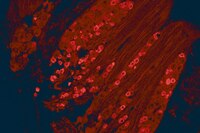
Characterization of bladder sensory neurons in the context of myelination, receptors for pain modulators, and acute responses to bladder inflammation.
Forrest, SL; Osborne, PB; Keast, JR
Frontiers in neuroscience
7
206
2013
Kivonat megmutatása
Bladder sensation is mediated by lumbosacral dorsal root ganglion neurons and is essential for normal voiding and nociception. Numerous electrophysiological, structural, and molecular changes occur in these neurons following inflammation. Defining which neurons undergo these changes is critical for understanding the mechanism underlying bladder pain and dysfunction. Our first aim was to define the chemical classes of bladder sensory neurons that express receptors for the endogenous modulators of nociceptor sensitivity, glial cell line-derived neurotrophic factor (GDNF), the related neurotrophic factor, artemin, and estrogens. Bladder sensory neurons of adult female Sprague-Dawley rats were identified with retrograde tracer. Diverse groups of neurons express these receptors, and some neurons express receptors for both neurotrophic factors and estrogens. Lumbar and sacral sensory neurons showed some distinct differences in their expression profile. We also distinguished the chemical profile of myelinated and unmyelinated bladder sensory neurons. Our second aim was to identify bladder sensory neurons likely to be undergoing structural remodeling during inflammation. Following systemic administration of cyclophosphamide (CYP), its renal metabolite acrolein causes transient urothelial loss, exposing local afferent terminals to a toxic environment. CYP induced expression of the injury-related immediate-early gene product, activating transcription factor-3 (ATF-3), in a small population of sacral nitrergic bladder sensory neurons. In conclusion, we have defined the bladder sensory neurons that express receptors for GDNF, artemin and estrogens. Our study has also identified a sub-population of sacral sensory neurons that are likely to be undergoing structural remodeling during acute inflammation of the bladder. Together these results contribute to increased understanding of the neurons that are known to be involved in pain modulation and hyperreflexia during inflammation. | | 24223534
 |
Transient receptor potential vanilloid type 1 channel (TRPV1) immunolocalization in the murine enteric nervous system is affected by the targeted C-terminal epitope of the applied antibody.
Buckinx, R; Van Nassauw, L; Avula, LR; Alpaerts, K; Adriaensen, D; Timmermans, JP
The journal of histochemistry and cytochemistry : official journal of the Histochemistry Society
61
421-32
2013
Kivonat megmutatása
The expression of transient receptor potential vanilloid type 1 channel (TRPV1) in the enteric nervous system is still the subject of debate. Although a number of studies have reported that TRPV1 is limited to extrinsic afferent fibers, other studies argue for an intrinsic expression of TRPV1. In the present study, reverse transcriptase PCR was employed to establish the expression of TRPV1 mRNA throughout the gastrointestinal tract. Using two antibodies directed against different epitopes of TRPV1, we were able to show at the protein level that the observed distribution pattern of TRPV1 is dependent on the antibody used in the immunohistochemical staining. A first antibody indeed mainly stained neuronal fibers, whereas a second antibody exclusively stained perikarya of enteric neurons throughout the mouse gastrointestinal tract. We argue that these different distribution patterns are due to the antibodies discriminating between different modulated forms of TRPV1 that influence the recognition of the targeted immunogen and as such distinguish intracellular from plasmalemmal forms of TRPV1. Our study is the first to directly compare these two antibodies within the same species and in identical conditions. Our observations underline that detailed knowledge of the epitope that is recognized by the antibodies employed in immunohistochemical procedures is a prerequisite for correctly interpreting experimental results. | Immunohistochemistry | 23482327
 |
Intrathecal injection of TRPV1 shRNA leads to increases in blood pressure in rats.
Yu, SQ; Wang, DH
Acta physiologica (Oxford, England)
203
139-47
2010
Kivonat megmutatása
The transient receptor potential vanilloid type 1 (TRPV1) channels have been implicated to play a role in blood pressure regulation. However, contribution of tissue specific TRPV1 to blood pressure regulation is largely unknown. Here, we test the hypothesis that TRPV1 expressed in dorsal root ganglia (DRG) of lower thoracic and upper lumbar segments (T8-L3) of the spinal cord and their central and peripheral terminals constitutes a counter regulatory mechanism preventing the increases in blood pressure.The expression of TRPV1 was knocked down by intrathecal injection of TRPV1 short-hairpin RNA (shRNA) in rats. Systolic blood pressure and mean arterial pressure (MAP) were recorded. The level of TRPV1 and tyrosine hydroxylase (TH) was measured by Western blot.Intrathecal injection of TRPV1 shRNA (6 μg kg(-1) day(-1) ) for 3 days increased systolic blood pressure and MAP when compared to rats that received control shRNA (control shRNA: 112 ± 2 vs. TRPV1 shRNA: 123 ± 2 mmHg). TRPV1 expression was suppressed in T8-L3 segments of dorsal horn and DRG as well as mesenteric arteries of rats given TRPV1 shRNA. Contents of TH, a marker of sympathetic nerves, were increased in mesenteric arteries of rats treated with TRPV1 shRNA. Pretreatment with the α1-adrenoceptor blocker, prazosin (1 mg kg(-1) day(-1) , p.o.), abolished the TRPV1 shRNA-induced pressor effects.Our data show that selective knockdown of TRPV1 expressed in DRG of T8-L3 segments of the spinal cord and their central and peripheral terminals increases blood pressure, suggesting that neuronal TRPV1 in these segments possesses a tonic anti-hypertensive effect possibly via suppression of the sympathetic nerve activity. | | 21518266
 |
Enhanced salt sensitivity following shRNA silencing of neuronal TRPV1 in rat spinal cord.
Yu, SQ; Wang, DH
Acta pharmacologica Sinica
32
845-52
2010
Kivonat megmutatása
To investigate the effects of selective knockdown of TRPV1 channels in the lower thoracic and upper lumbar segments of spinal cord, dorsal root ganglia (DRG) and mesenteric arteries on rat blood pressure responses to high salt intake.TRPV1 short-hairpin RNA (shRNA) was delivered using intrathecal injection (6 μg · kg(-1) · d(-1), for 3 d). Levels of TRPV1 and tyrosine hydroxylase expression were determined by Western blot analysis. Systolic blood pressure and mean arterial pressure (MAP) were examined using tail-cuff and direct arterial measurement, respectively.In rats injected with control shRNA, high-salt diet (HS) caused higher systolic blood pressure compared with normal-salt diet (NS) (HS:149 ± 4 mmHg; NS:126 ± 2 mmHg, Pless than 0.05). Intrathecal injection of TRPV1 shRNA significantly increased the systolic blood pressure in both HS rats and NS rats (HS:169 ± 3 mmHg; NS:139 ± 2 mmHg). The increases was greater in HS rats than in NS rats (HS: 13.9% ± 1.8%; NS: 9.8 ± 0.7, Pless than 0.05). After TRPV1 shRNA treatment, TRPV1 expression in the dorsal horn and DRG of T8-L3 segments and in mesenteric arteries was knocked down to a greater extent in HS rats compared with NS rats. Blockade of α1-adrenoceptors abolished the TRPV1 shRNA-induced pressor effects. In rats injected with TRPV1 shRNA, level of tyrosine hydroxylase in mesenteric arteries was increased to a greater extent in HS rats compared with NS rats.Selective knockdown of TRPV1 expression in the lower thoracic and upper lumbar segments of spinal cord, DRG, and mesenteric arteries enhanced the prohypertensive effects of high salt intake, suggesting that TRPV1 channels in these sites protect against increased salt sensitivity, possibly via suppression of sympatho-excitatory responses. | | 21642952
 |
Protein kinase D isoforms are expressed in rat and mouse primary sensory neurons and are activated by agonists of protease-activated receptor 2.
Amadesi S, Grant AD, Cottrell GS, Vaksman N, Poole DP, Rozengurt E, Bunnett NW
J Comp Neurol
516
141-56
2009
Kivonat megmutatása
Serine proteases generated during injury and inflammation cleave protease-activated receptor 2 (PAR(2)) on primary sensory neurons to induce neurogenic inflammation and hyperalgesia. Hyperalgesia requires sensitization of transient receptor potential vanilloid (TRPV) ion channels by mechanisms involving phospholipase C and protein kinase C (PKC). The protein kinase D (PKD) serine/threonine kinases are activated by diacylglycerol and PKCs and can phosphorylate TRPV1. Thus, PKDs may participate in novel signal transduction pathways triggered by serine proteases during inflammation and pain. However, it is not known whether PAR(2) activates PKD, and the expression of PKD isoforms by nociceptive neurons is poorly characterized. By using HEK293 cells transfected with PKDs, we found that PAR(2) stimulation promoted plasma membrane translocation and phosphorylation of PKD1, PKD2, and PKD3, indicating activation. This effect was partially dependent on PKCepsilon. By immunofluorescence and confocal microscopy, with antibodies against PKD1/PKD2 and PKD3 and neuronal markers, we found that PKDs were expressed in rat and mouse dorsal root ganglia (DRG) neurons, including nociceptive neurons that expressed TRPV1, PAR(2), and neuropeptides. PAR(2) agonist induced phosphorylation of PKD in cultured DRG neurons, indicating PKD activation. Intraplantar injection of PAR(2) agonist also caused phosphorylation of PKD in neurons of lumbar DRG, confirming activation in vivo. Thus, PKD1, PKD2, and PKD3 are expressed in primary sensory neurons that mediate neurogenic inflammation and pain transmission, and PAR(2) agonists activate PKDs in HEK293 cells and DRG neurons in culture and in intact animals. PKD may be a novel component of a signal transduction pathway for protease-induced activation of nociceptive neurons and an important new target for antiinflammatory and analgesic therapies. Copyright 2009 Wiley-Liss, Inc. Teljes cikk | | 19575452
 |
The role of transient receptor potential vanilloid 1 in mechanical and chemical visceral hyperalgesia following experimental colitis.
A Miranda,E Nordstrom,A Mannem,C Smith,B Banerjee,J N Sengupta
Neuroscience
148
2007
Kivonat megmutatása
The transient receptor potential vanilloid 1 receptor (TRPV1) is an important nociceptor involved in neurogenic inflammation. We aimed to examine the role of TRPV1 in experimental colitis and in the development of visceral hypersensitivity to mechanical and chemical stimulation. Male Sprague-Dawley rats received a single dose of trinitrobenzenesulfonic acid (TNBS) in the distal colon. In the preemptive group, rats received the TRPV1 receptor antagonist JYL1421 (10 mumol/kg, i.v.) or vehicle 15 min prior to TNBS followed by daily doses for 7 days. In the post-inflammation group, rats received JYL1421 daily for 7 days starting on day 7 following TNBS. The visceromotor response (VMR) to colorectal distension (CRD), intraluminal capsaicin, capsaicin vehicle (pH 6.7) or acidic saline (pH 5.0) was assessed in all groups and compared with controls and naïve rats. Colon inflammation was evaluated with H&E staining and myeloperoxidase (MPO) activity. TRPV1 immunoreactivity was assessed in the thoraco-lumbar (TL) and lumbo-sacral (LS) dorsal root ganglia (DRG) neurons. In the preemptive vehicle group, TNBS resulted in a significant increase in the VMR to CRD, intraluminal capsaicin and acidic saline compared the JYL1421-treated group (P<0.05). Absence of microscopic colitis and significantly reduced MPO activity was also evident compared with vehicle-treated rats (P<0.05). TRPV1 immunoreactivity in the TL (69.1+/-4.6%) and LS (66.4+/-4.2%) DRG in vehicle-treated rats was increased following TNBS but significantly lower in the preemptive JYL1421-treated group (28.6+/-3.9 and 32.3+/-2.3 respectively, P<0.05). JYL1421 in the post-inflammation group improved microscopic colitis and significantly decreased the VMR to CRD compared with vehicle (P<0.05, >/=30 mm Hg) but had no effect on the VMR to chemical stimulation. TRPV1 immunoreactivity in the TL and LS DRG was no different from vehicle or naïve controls. These results suggest an important role for TRPV1 channel in the development of inflammation and subsequent mechanical and chemical visceral hyperalgesia. Teljes cikk | | 17719181
 |
Activation of vanilloid receptor type I in the endoplasmic reticulum fails to activate store-operated Ca2+ entry
Wisnoskey, B. J. et al.
Biochem. J. , 372(Pt 2):517-528 (2003)
2003
| | 12608892
 |
Topography of the vanilloid receptor in the human bladder: more than just the nerve fibers.
Dieter Ost, Tania Roskams, Frank Van Der Aa, Dirk De Ridder
The Journal of urology
168
293-7
2002
Kivonat megmutatása
PURPOSE: We determined the presence and distribution of vanilloid receptor-1 in the human bladder and confirmed or rejected previous findings of other groups that used indirect methods or vanilloid receptor-1 antibodies made by immunizing experimental animals. Also, we tested the reproducibility of results using commercially available antibodies against the N-terminus and C-terminus of the vanilloid receptor. MATERIALS AND METHODS: A total of 11 normal bladder tissue samples were obtained from cystectomy specimens and fresh frozen processed. Specimens were studied by immunohistochemistry and confocal laser microscopy using 3 vanilloid receptor-1 antibodies. Immunohistochemical co-localization studies for neurofilament, neuronal nitric oxide synthase and nerve growth factor were performed. RESULTS: Our results confirm the presence of vanilloid receptor-1 on nonmyelinated and myelinated nerve fibers. There was vanilloid receptor-1 immunoreactivity on smooth muscle cells but different sensitivities for the antibodies. There was immunoreactivity on interstitial cells located in the suburothelium and intermuscular septa of the muscularis. There was co-localization of neuronal nitric oxide synthase with interstitial cells but not with neurofilament. No co-localization was found for nerve growth factor and vanilloid receptor-1. CONCLUSIONS: Vanilloid receptor-1 is located on small unmyelinated and myelinated nerve fibers. In addition, vanilloid receptor-1 is also present on interstitial cells in the suburothelium. There is smooth muscle cell immunoreactivity but a difference in antibodies raised against the C-terminus and N-terminus. These data suggest that the current hypothesis about the mechanism of action of vanilloids is through blocking the afferent reflex arc must be revised and the function of interstitial cells deserves further attention. | | 12050559
 |
Immunocytochemical localization of the vanilloid receptor 1 (VR1): relationship to neuropeptides, the P2X3 purinoceptor and IB4 binding sites
Guo, A. et al.
Eur. J. Neurosci., 11(3):946-958 (1999)
1998
| | 10103088
 |


