Millipore Sigma Vibrant Logo
Attention: We have moved. Merck Millipore products are no longer available for purchase on MerckMillipore.com.Learn More

MABN694 Anti-CHD3 Antibody, clone 2G4

MABN694
100 µL  
Purchase on Sigma-Aldrich

Különleges ajánlatok

Áttekintés

Replacement Information

Különleges ajánlatok

Kulcsspecifikációk táblázata

Species ReactivityKey ApplicationsHostFormatAntibody Type
H, MWB, IHC, IF, FCMAscitesMonoclonal Antibody
Description
Catalogue NumberMABN694
ReplacesMABE482
DescriptionAnti-CHD3 Antibody, clone 2G4
Alternate Names
  • Chromodomain-helicase-DNA-binding protein 3
  • ATP-dependent helicase CHD3
  • Mi-2 autoantigen 240 kDa protein
  • Mi2-alpha
  • Zinc finger helicase
  • hZFH
  • CHD3
Background InformationChromodomain-helicase-DNA-binding protein 3 (CHD-3) or alternatively known as ATP-dependent helicase CHD3, Mi-2 autoantigen 240kDa protein, Mi2-alpha, Zinc finger helicase (hZFH) and encoded by the human gene CHD3 is a member of the CHD family of proteins which are characterized by the presence of a chromo (chromatin organization modifier) domains and SNF2-related helicase/ATPase domains. CHD3 is one member of a large histone deacetylase complex called NuRD. CHD3 is one of the ATPases that form part of the NuRD complex. The NuRD complex operates in the remodeling of chromatin by deacetylating histones and thus typically functions as a transcriptional repressor, though NuRD can also act as an activator depending upon the system studied. CHD3 via the NuRD complex also interacts with HDAC1 and HDAC2. CHD3 is also required for anchoring of the centrosomal protein pericentrin and for the organization of the spindle and centrosome during cell division. It is typically located in the nucleus but CHD3 is seen cytoplasmically during mitosis associated with the centrosomes. CHD3 is expressed by most cells and tissues. Interestingly CHD3 is also a hallmark auto antigen of MI-2 dermatomyositis. EMD-Millipore’s Anti-CHD3 mouse monoclonal antibody has been tested in western blot against HeLa, K562, Jurkat, NTERA-2, HEK293, Raji cells lysates and mouse brain tissue lysate and in paraffin embedded immunohistochemistry on human colon cancer tissue sections. The antibody has also been successfully tested in fluorescent immunocytochemistry on HeLa cells in culture and by flow cytometry on K562 cells in culture.
References
Product Information
FormatAscites
Control
  • mouse brain, HeLa, K562, Jurkat, NTERA-2, HEK293, Raji lysates
PresentationMouse monoclonal IgG1 ascitic fluid containing up to 0.1% sodium azide.
Quality LevelMQ100
Applications
ApplicationDetect CHD-3 using this mouse monoclonal antibody, Anti-CHD3 Antibody, clone 2G4 validated for use in western blotting, IHC, Immunofluorescence & Flow Cytometry.
Key Applications
  • Western Blotting
  • Immunohistochemistry
  • Immunofluorescence
  • Flow Cytometry
Application NotesImmunohistochemistry Analysis: A 1:200-1,000 dilution from a representative lot detected CHD3 in colon cancer tissue.

Immunofluorescence Analysis: A 1:200-1,000 dilution from a representative lot detected CHD3 in HeLa cells.

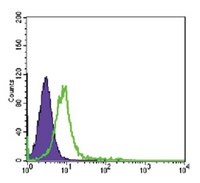
Flow cytometry analysis: A 1:200-400 dilution from a representative lot detected CHD3 in K562 cells.

Optimal working dilutions must be determined by end user.
Biological Information
ImmunogenPurified recombinant fragment of human CHD3 expressed in E. Coli.
Clone2G4
HostMouse
IsotypeIgG1
Species Reactivity
  • Human
  • Mouse
Antibody TypeMonoclonal Antibody
Gene Symbol
  • ZFH
  • Mi-2a
  • Mi2-ALPHA
  • CHD3
Purification MethodUnpurified
UniProt Number
Molecular Weight~220 kDa observed. Uncharacterized bands may appear in some lysate(s).
Physicochemical Information
Dimensions
Materials Information
Toxicological Information
Safety Information according to GHS
Safety Information
Product Usage Statements
Quality AssuranceEvaluated by Western Blotting in mouse brain, HeLa, K562, Jurkat, NTERA-2, HEK293, Raji lysates.

Western Blotting Analysis: A 1:500-2,000 dilution of this antibody detected CHD3 in mouse brain, HeLa, K562, Jurkat, NTERA-2, HEK293, Raji lysates.
Usage Statement
  • Unless otherwise stated in our catalog or other company documentation accompanying the product(s), our products are intended for research use only and are not to be used for any other purpose, which includes but is not limited to, unauthorized commercial uses, in vitro diagnostic uses, ex vivo or in vivo therapeutic uses or any type of consumption or application to humans or animals.
Storage and Shipping Information
Storage ConditionsStable for 1 year at -20°C from date of receipt.
Handling Recommendations: Upon receipt and prior to removing the cap, centrifuge the vial and gently mix the solution. Aliquot into microcentrifuge tubes and store at -20°C. Avoid repeated freeze/thaw cycles, which may damage IgG and affect product performance.
Packaging Information
Material Size100 µL
Transport Information
Supplemental Information
Specifications
Global Trade Item Number
Katalógusszám GTIN
MABN694 04053252920745