Human leukocyte antigen-G is frequently expressed in glioblastoma and may be induced in vitro by combined 5-aza-2'-deoxycytidine and interferon-γ treatments: results from a multicentric study.
Wastowski, IJ; Simões, RT; Yaghi, L; Donadi, EA; Pancoto, JT; Poras, I; Lechapt-Zalcman, E; Bernaudin, M; Valable, S; Carlotti, CG; Flajollet, S; Jensen, SS; Ferrone, S; Carosella, ED; Kristensen, BW; Moreau, P
The American journal of pathology
182
540-52
2013
显示摘要
Human leukocyte antigen-G (HLA-G) is a nonclassical major histocompatibility complex (MHC) class I molecule involved in immune tolerance processes, playing an important role in the maintenance of the semi-allogeneic fetus. Although HLA-G expression is restricted in normal tissues, it is broadly expressed in malignant tumors and may favor tumor immune escape. We analyzed HLA-G protein and mRNA expression in tumor samples from patients with glioblastoma collected in France, Denmark, and Brazil. We found HLA-G protein expression in 65 of 108 samples and mRNA in 20 of 21 samples. The absence of HLA-G protein expression was associated with a better long-term survival rate. The mechanisms underlying HLA-G gene expression were investigated in glioma cell lines U251MG, D247MG, and U138MG. Induction of HLA-G transcriptional activity was dependent of 5-aza-2'-deoxycytidine treatment and enhanced by interferon-γ. HLA-G protein expression was observed in U251MG cells only. These cells exhibited a permissive chromatin state at the HLA-G gene promoter and the highest levels of induced HLA-G transcriptional activity following 5-aza-2'-deoxycytidine treatment. Several antigen-presenting machinery components were up-regulated in U251MG cells after demethylating and IFN-γ treatments, suggesting an effect on the up-regulation of HLA-G cell surface expression. Therefore, because of its role in tumor tolerance, HLA-G found to be expressed in glioblastoma samples should be taken into consideration in clinical studies on the pathology and in the design of therapeutic strategies to prevent its expression in HLA-G-negative tumors. | | 23219427
 |
Repeated social defeat selectively increases δFosB expression and histone H3 acetylation in the infralimbic medial prefrontal cortex.
Hinwood, M; Tynan, RJ; Day, TA; Walker, FR
Cerebral cortex (New York, N.Y. : 1991)
21
262-71
2011
显示摘要
Exposure to social stress has been linked to the development and maintenance of mood-related psychopathology; however, the underlying neurobiological changes remain uncertain. In this study, we examined numbers of δFosB-immunoreactive cells in the forebrains of rats subjected to 12 episodes of social defeat. This was achieved using the social conflict model whereby animals are introduced into the home cage of older males ("residents") trained to attack and defeat all such "intruders"; importantly, controls were treated identically except that the resident was absent. Our results indicated that the only region in which δFosB-positive cells were found in significantly higher numbers in intruders than in controls was the infralimbic medial prefrontal cortex (mPFC). This same effect was not apparent using another psychological stressor, noise stress. Cells of the infralimbic mPFC also displayed evidence of chromatin remodeling. We found that exposure to repeated episodes of social defeat increased numbers of cells immunoreactive for histone H3 acetylation, but not for histone H3 phosphoacetylation, in the infralimbic mPFC. Collectively, these findings highlight the importance of the infralimbic mPFC in responding to social stress-a finding that provides insight into the possible neurobiological alterations associated with stress-induced psychiatric illness. | | 20513656
 |
Epigenetic regulation of a murine retrotransposon by a dual histone modification mark.
Brunmeir, R; Lagger, S; Simboeck, E; Sawicka, A; Egger, G; Hagelkruys, A; Zhang, Y; Matthias, P; Miller, WJ; Seiser, C
PLoS genetics
6
e1000927
2010
显示摘要
Large fractions of eukaryotic genomes contain repetitive sequences of which the vast majority is derived from transposable elements (TEs). In order to inactivate those potentially harmful elements, host organisms silence TEs via methylation of transposon DNA and packaging into chromatin associated with repressive histone marks. The contribution of individual histone modifications in this process is not completely resolved. Therefore, we aimed to define the role of reversible histone acetylation, a modification commonly associated with transcriptional activity, in transcriptional regulation of murine TEs. We surveyed histone acetylation patterns and expression levels of ten different murine TEs in mouse fibroblasts with altered histone acetylation levels, which was achieved via chemical HDAC inhibition with trichostatin A (TSA), or genetic inactivation of the major deacetylase HDAC1. We found that one LTR retrotransposon family encompassing virus-like 30S elements (VL30) showed significant histone H3 hyperacetylation and strong transcriptional activation in response to TSA treatment. Analysis of VL30 transcripts revealed that increased VL30 transcription is due to enhanced expression of a limited number of genomic elements, with one locus being particularly responsive to HDAC inhibition. Importantly, transcriptional induction of VL30 was entirely dependent on the activation of MAP kinase pathways, resulting in serine 10 phosphorylation at histone H3. Stimulation of MAP kinase cascades together with HDAC inhibition led to simultaneous phosphorylation and acetylation (phosphoacetylation) of histone H3 at the VL30 regulatory region. The presence of the phosphoacetylation mark at VL30 LTRs was linked with full transcriptional activation of the mobile element. Our data indicate that the activity of different TEs is controlled by distinct chromatin modifications. We show that activation of a specific mobile element is linked to a dual epigenetic mark and propose a model whereby phosphoacetylation of histone H3 is crucial for full transcriptional activation of VL30 elements. | Western Blotting | 20442873
 |
14-3-3 mediates histone cross-talk during transcription elongation in Drosophila.
Karam, CS; Kellner, WA; Takenaka, N; Clemmons, AW; Corces, VG
PLoS genetics
6
e1000975
2010
显示摘要
Post-translational modifications of histone proteins modulate the binding of transcription regulators to chromatin. Studies in Drosophila have shown that the phosphorylation of histone H3 at Ser10 (H3S10ph) by JIL-1 is required specifically during early transcription elongation. 14-3-3 proteins bind H3 only when phosphorylated, providing mechanistic insights into the role of H3S10ph in transcription. Findings presented here show that 14-3-3 functions downstream of H3S10ph during transcription elongation. 14-3-3 proteins localize to active genes in a JIL-1-dependent manner. In the absence of 14-3-3, levels of actively elongating RNA polymerase II are severely diminished. 14-3-3 proteins interact with Elongator protein 3 (Elp3), an acetyltransferase that functions during transcription elongation. JIL-1 and 14-3-3 are required for Elp3 binding to chromatin, and in the absence of either protein, levels of H3K9 acetylation are significantly reduced. These results suggest that 14-3-3 proteins mediate cross-talk between histone phosphorylation and acetylation at a critical step in transcription elongation. 全文本文章 | Western Blotting | 20532201
 |
Lovastatin-induced cholesterol depletion affects both apical sorting and endocytosis of aquaporin-2 in renal cells.
Procino G, Barbieri C, Carmosino M, Rizzo F, Valenti G, Svelto M
American journal of physiology. Renal physiology
298
F266-78. Epub 2009 Nov 18.
2010
显示摘要
Vasopressin causes the redistribution of the water channel aquaporin-2 (AQP2) from cytoplasmic storage vesicles to the apical plasma membrane of collecting duct principal cells, leading to urine concentration. The molecular mechanisms regulating the selective apical sorting of AQP2 are only partially uncovered. In this work, we investigate whether AQP2 sorting/trafficking is regulated by its association with membrane rafts. In both MCD4 cells and rat kidney, AQP2 preferentially associated with Lubrol WX-insoluble membranes regardless of its presence in the storage compartment or at the apical membrane. Block-and-release experiments indicate that 1) AQP2 associates with detergent-resistant membranes early in the biosynthetic pathway; 2) strong cholesterol depletion delays the exit of AQP2 from the trans-Golgi network. Interestingly, mild cholesterol depletion promoted a dramatic accumulation of AQP2 at the apical plasma membrane in MCD4 cells in the absence of forskolin stimulation. An internalization assay showed that AQP2 endocytosis was clearly reduced under this experimental condition. Taken together, these data suggest that association with membrane rafts may regulate both AQP2 apical sorting and endocytosis. | | 19923410
 |
Multiple chromatin-bound protein kinases assemble factors that regulate insulin gene transcription.
Lawrence, MC; Shao, C; McGlynn, K; Naziruddin, B; Levy, MF; Cobb, MH
Proceedings of the National Academy of Sciences of the United States of America
106
22181-6
2009
显示摘要
During the onset of diabetes, pancreatic beta cells become unable to produce sufficient insulin to maintain blood glucose within the normal range. Proinflammatory cytokines have been implicated in impaired beta cell function. To understand more about the molecular events that reduce insulin gene transcription, we examined the effects of hyperglycemia alone and together with the proinflammatory cytokine interleukin-1beta (IL-1beta) on signal transduction pathways that regulate insulin gene transcription. Exposure to IL-1beta in fasting glucose activated multiple protein kinases that associate with the insulin gene promoter and transiently increased insulin gene transcription in beta cells. In contrast, cells exposed to hyperglycemic conditions were sensitized to the inhibitory actions of IL-1beta. Under these conditions, IL-1beta caused the association of the same protein kinases, but a different combination of transcription factors with the insulin gene promoter and began to reduce transcription within 2 h; stimulatory factors were lost, RNA polymerase II was lost, and inhibitory factors were bound to the promoter in a kinase-dependent manner. | | 20018749
 |
Drug-induced activation of dopamine D(1) receptor signaling and inhibition of class I/II histone deacetylase induce chromatin remodeling in reward circuitry and modulate cocaine-related behaviors.
Schroeder, FA; Penta, KL; Matevossian, A; Jones, SR; Konradi, C; Tapper, AR; Akbarian, S
Neuropsychopharmacology : official publication of the American College of Neuropsychopharmacology
33
2981-92
2008
显示摘要
Chromatin remodeling, including histone modification, is involved in stimulant-induced gene expression and addiction behavior. To further explore the role of dopamine D(1) receptor signaling, we measured cocaine-related locomotor activity and place preference in mice pretreated for up to 10 days with the D(1) agonist SKF82958 and/or the histone deacetylase inhibitor (HDACi), sodium butyrate. Cotreatment with D(1) agonist and HDACi significantly enhanced cocaine-induced locomotor activity and place preference, in comparison to single-drug regimens. However, butyrate-mediated reward effects were transient and only apparent within 2 days after the last HDACi treatment. These behavioral changes were associated with histone modification changes in striatum and ventral midbrain: (1) a generalized increase in H3 phosphoacetylation in striatal neurons was dependent on activation of D(1) receptors; (2) H3 deacetylation at promoter sequences of tyrosine hydroxylase (Th) and brain-derived neurotrophic factor (Bdnf) in ventral midbrain, together with upregulation of the corresponding gene transcripts after cotreatment with D(1) agonist and HDACi. Collectively, these findings imply that D(1) receptor-regulated histone (phospho)acetylation and gene expression in reward circuitry is differentially regulated in a region-specific manner. Given that the combination of D(1) agonist and HDACi enhances cocaine-related sensitization and reward, the therapeutic benefits of D(1) receptor antagonists and histone acetyl-transferase inhibitors (HATi) warrant further investigation in experimental models of stimulant abuse. | | 18288092
 |
Distinct roles of the steroid receptor coactivator 1 and of MED1 in retinoid-induced transcription and cellular differentiation.
Flajollet, S; Lefebvre, B; Rachez, C; Lefebvre, P
The Journal of biological chemistry
281
20338-48
2006
显示摘要
Retinoic acid receptors (RARs) are the molecular relays of retinoid action on transcription, cellular differentiation and apoptosis. Transcriptional activation of retinoid-regulated promoters requires the dismissal of corepressors and the recruitment of coactivators to promoter-bound RAR. RARs recruit in vitro a plethora of coactivators whose actual contribution to retinoid-induced transcription is poorly characterized in vivo. Embryonal carcinoma P19 cells, which are highly sensitive to retinoids, were depleted from archetypical coactivators by RNAi. SRC1-deficient P19 cells showed severely compromised retinoid-induced responses, in agreement with the supposed role of SRC1 as a RAR coactivator. Unexpectedly, Med1/TRAP220/DRIP205-depleted cells exhibited an exacerbated response to retinoids, both in terms transcriptional responses and of cellular differentiation. Med1 depletion affected TFIIH and cdk9 detection at the prototypical retinoid-regulated RARbeta2 promoter, and favored a higher RNA polymerase II detection in transcribed regions of the RARbeta2 gene. Furthermore, the nature of the ligand impacted strongly on the ability of RARs to interact with a given coactivator and to activate transcription in intact cells. Thus RAR accomplishes transcriptional activation as a function of the ligand structure, by recruiting regulatory complexes which control distinct molecular events at retinoid-regulated promoters. | | 16723356
 |
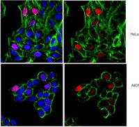
Inhibition of mixed-lineage kinase (MLK) activity during G2-phase disrupts microtubule formation and mitotic progression in HeLa cells.
Hyukjin Cha, Surabhi Dangi, Carolyn E Machamer, Paul Shapiro
Cellular signalling
18
93-104
2006
显示摘要
The mixed-lineage kinases (MLK) are serine/threonine protein kinases that regulate mitogen-activated protein (MAP) kinase signaling pathways in response to extracellular signals. Recent studies indicate that MLK activity may promote neuronal cell death through activation of the c-Jun NH2-terminal kinase (JNK) family of MAP kinases. Thus, inhibitors of MLK activity may be clinically useful for delaying the progression of neurodegenerative diseases, such as Parkinson's. In proliferating non-neuronal cells, MLK may have the opposite effect of promoting cell proliferation. In the current studies we examined the requirement for MLK proteins in regulating cell proliferation by examining MLK function during G2 and M-phase of the cell cycle. The MLK inhibitor CEP-11004 prevented HeLa cell proliferation by delaying mitotic progression. Closer examination revealed that HeLa cells treated with CEP-11004 during G2-phase entered mitosis similar to untreated G2-phase cells. However, CEP-11004 treated cells failed to properly exit mitosis and arrested in a pro-metaphase state. Partial reversal of the CEP-11004 induced mitotic arrest could be achieved by overexpression of exogenous MLK3. The effects of CEP-11004 treatment on mitotic events included the inhibition of histone H3 phosphorylation during prophase and prior to nuclear envelope breakdown and the formation of aberrant mitotic spindles. These data indicate that MLK3 might be a unique target to selectively inhibit transformed cell proliferation by disrupting mitotic spindle formation resulting in mitotic arrest. | | 15923109
 |
Targeted expression of cyclin D2 results in cardiomyocyte DNA synthesis and infarct regression in transgenic mice.
Pasumarthi, KB; Nakajima, H; Nakajima, HO; Soonpaa, MH; Field, LJ
Circulation research
96
110-8
2005
显示摘要
Restriction point transit and commitment to a new round of cell division is regulated by the activity of cyclin-dependent kinase 4 and its obligate activating partners, the D-type cyclins. In this study, we examined the ability of D-type cyclins to promote cardiomyocyte cell cycle activity. Adult transgenic mice expressing cyclin D1, D2, or D3 under the regulation of the alpha cardiac myosin heavy chain promoter exhibited high rates of cardiomyocyte DNA synthesis under baseline conditions. Cardiac injury in mice expressing cyclin D1 or D3 resulted in cytoplasmic cyclin D accumulation, with a concomitant reduction in the level of cardiomyocyte DNA synthesis. In contrast, cardiac injury in mice expressing cyclin D2 did not alter subcellular cyclin localization. Consequently, cardiomyocyte cell cycle activity persisted in injured hearts expressing cyclin D2, ultimately resulting in infarct regression. These data suggested that modulation of D-type cyclins could be exploited to promote regenerative growth in injured hearts. | Immunohistochemistry | 15576649
 |


