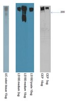
mAb Das-1 is specific for high-risk and malignant intraductal papillary mucinous neoplasm (IPMN).
Das, KK; Xiao, H; Geng, X; Fernandez-Del-Castillo, C; Morales-Oyarvide, V; Daglilar, E; Forcione, DG; Bounds, BC; Brugge, WR; Pitman, MB; Mino-Kenudson, M; Das, KM
Gut
63
1626-34
2014
显示摘要
Intraductal papillary mucinous neoplasm (IPMN) consists of four epithelial subtypes that correlate with histological grades and risks for malignant transformation. mAb Das-1 is a monoclonal antibody against a colonic epithelial phenotype that is reactive to premalignant conditions of the upper GI tract. We sought to assess the ability of mAb Das-1 to identify IPMN with high risk of malignant transformation.mAb Das-1 reactivity was evaluated in 94 patients with IPMNs by immunohistochemistry. Lesional fluid from 38 separate patients with IPMN (n=27), low-grade non-mucinous cystic neoplasms (n=7) and pseudocysts (n=4) was analysed by ELISA and western blot.Immunohistochemistry-Normal pancreatic ducts were non-reactive and low-grade gastric-type IPMN (IPMN-G) (1/17) and intermediate-grade IPMN-G (1/23) were minimally reactive with mAb Das-1. In contrast, mAb Das-1 reactivity was significantly higher in high-risk/malignant lesions (p<0.0001) including: intestinal-type IPMN with intermediate-grade dysplasia (9/10); high-grade dysplasia of gastric (4/7), intestinal (12/12), oncocytic (2/2) and pancreatobiliary types (2/2); and invasive tubular (8/12), colloid (7/7) and oncocytic (2/2) carcinoma. The sensitivity and specificity of mAb Das-1 for high-risk/malignant IPMNs were 85% and 95%, respectively. Lesional fluid-Samples from low- and intermediate-grade IPMN-G (n=9), and other low-grade/benign non-mucinous lesions demonstrated little reactivity with mAb Das-1. Conversely, cyst fluid from high-risk/malignant IPMNs (n=18) expressed significantly higher reactivity (p<0.0001). The sensitivity and specificity of mAbDas-1 in detecting high-risk/malignant IPMNs were 89% and 100%, respectively.mAb Das-1 reacts with high specificity to tissue and cyst fluid from high-risk/malignant IPMNs and thus may help in preoperative clinical risk stratification. | 24277729
 |
Effects of Helicobacter pylori infection on genetic instability, the aberrant CpG island methylation status and the cellular phenotype in Barrett's esophagus in a Japanese population.
Moriichi, K; Watari, J; Das, KM; Tanabe, H; Fujiya, M; Ashida, T; Kohgo, Y
International journal of cancer. Journal international du cancer
124
1263-9
2009
显示摘要
Genetic or epigenetic alterations in Barrett's esophagus (BE) with/without Helicobacter pylori (H. pylori) infection remain unclear. We examined the effects of H. pylori infection on genetic instability (GIN), the CpG island methylation status and a biomarker related to BE carcinogenesis. We analyzed 113 Japanese individuals with endoscopically suspected BE. The patients included, Group CLE (n = 25): no specialized intestinal metaplasia (SIM) in a columnar lined epithelium (control); Group BE (n = 88): all had SIM. Microsatellite instability and a loss of heterozygosity as GIN, the methylation status at hMLH1, E-cadherin, p16 and APC, and immunoreactivity using a monoclonal antibody (mAb) Das-1, which specifically reacts with BE, were evaluated. Nine additional patients with BE were prospectively followed up for 2 years after successful H. pylori eradication. The frequency of GIN, methylation at E-cadherin and APC, and mAb Das-1 reactivity in Group BE was significantly higher than that in Group CLE (p < 0.0001, p < 0.0001 and p < 0.005, and p < 0.0001, respectively). Furthermore, GIN, E-cadherin methylation and mAb Das-1 reactivity showed a significantly higher incidence in patients with H.pylori infection than in those without H. pylori infection (p < 0.01, p < 0.005, and p < 0.01, respectively). Interestingly, the patients from Group BE were observed to change to a stable state of molecular alterations in 60% for GIN, 42.9% for E-cadherin methylation and 55.6% for APC methylation, or a reduction of mAb Das-1 reactivity was noted in 25% following eradication. H. pylori infection may therefore affect these molecular alterations associated with the pathogenesis of BE, to some degree, in the Japanese population. | 19048617
 |
Phenotypic differences between esophageal and gastric intestinal metaplasia.
Piazuelo, MB; Haque, S; Delgado, A; Du, JX; Rodriguez, F; Correa, P
Modern pathology : an official journal of the United States and Canadian Academy of Pathology, Inc
17
62-74
2004
显示摘要
Intestinal metaplasia is a cancer precursor in the esophagus and the stomach. Marked differences exist between the carcinogenic processes in the two locations in terms of natural history and clinical significance. We investigated biopsies from 52 patients with Barrett's esophagus and from 50 patients with gastric intestinal metaplasia in an attempt to throw light on their pathogenic processes. Morphologic characteristics, presence of Helicobacter pylori (H. pylori), and markers of differentiation, inflammation, and proliferation were evaluated by histochemical and immunohistochemical techniques. The area covered by incomplete type of intestinal metaplasia and the proportion of sulfomucins were significantly higher in the esophagus than in the stomach. Immunoreactivity with MUC1, MUC2, MUC5AC, Das-1, cytokeratins 7 and 20, inducible nitric oxide synthase and cyclooxygenase-2 antibodies was also significantly greater in Barrett's esophagus than in gastric intestinal metaplasia. In gastric intestinal metaplasia, the presence of MUC1, MUC5AC, Das-1 and cytokeratin 7 was restricted to areas with the incomplete type of metaplasia. Cell proliferation (Ki-67) was significantly higher in Barrett's esophagus than in gastric intestinal metaplasia. H. pylori was absent in all of the patients with Barrett's esophagus, while it was present in 70% of the patients with gastric intestinal metaplasia. Our observations made clear that Barrett's esophagus shares some phenotypic characteristics with gastric intestinal metaplasia, leading us to suggest that both could arise in response to injuries with eventual carcinogenic potential. However, the progression to more advanced lesions could be modulated by the nature of the carcinogenic insult. | 14631367
 |
Gastric intestinal metaplasia as detected by a monoclonal antibody is highly associated with gastric adenocarcinoma.
Mirza, ZK; Das, KK; Slate, J; Mapitigama, RN; Amenta, PS; Griffel, LH; Ramsundar, L; Watari, J; Yokota, K; Tanabe, H; Sato, T; Kohgo, Y; Das, KM
Gut
52
807-12
2003
显示摘要
Some forms of gastric intestinal metaplasia (GIM) may be precancerous but the cellular phenotype that predisposes to gastric carcinogenesis is not well characterised. Mucin staining, as a means of differentiating GIM, is difficult. A monoclonal antibody, mAb Das-1 (initially called 7E(12)H(12)), whose staining is phenotypically specific to colon epithelium, was used to investigate this issue.Using mAb Das-1, by a sensitive immunoperoxidase assay, we examined histologically confirmed GIM specimens from two countries, the USA and Japan. A total of 150 patients comprised three groups: group A, GIM (fields away from the cancer area) from patients with gastric carcinoma (n=60); group B, GIM with chronic gastritis (without gastric carcinoma) (n=72); and group C, chronic gastritis without GIM (n=18).Fifty six of 60 (93%) patients with GIM (both goblet and non-goblet metaplastic cells) from group A reacted intensely with mAb Das-1. Cancer areas from the same 56 patients also reacted. In contrast, 25/72 (35%) samples of GIM from patients in group B reacted with mAb Das-1 (group A v B, p<0.0001). None of the samples from group C reacted with the mAb.Reactivity of mAb Das-1 is clinically useful to simplify and differentiate the phenotypes of GIM. The colonic phenotype of GIM, as identified by mAb Das-1, is strongly associated with gastric carcinoma. | 12740335
 |
Phenotype of Barrett's esophagus and intestinal metaplasia of the distal esophagus and gastroesophageal junction: an immunohistochemical study of cytokeratins 7 and 20, Das-1 and 45 MI.
Glickman, JN; Wang, H; Das, KM; Goyal, RK; Spechler, SJ; Antonioli, D; Odze, RD
The American journal of surgical pathology
25
87-94
2001
显示摘要
The pathogenesis of short segment Barrett's esophagus (SSBE) and intestinal metaplasia (IM) of the gastroesophageal junction (IMGEJ) are poorly understood. Also, these conditions are difficult to distinguish from one another based solely on endoscopic and pathologic criteria. Therefore, the aim of this study was to evaluate the immunophenotypic features of SSBE and IMGEJ and to compare the results with lesions of known etiologies: long segment BE (LSBE) caused by reflux disease and Helicobacter pylori-induced IM of the gastric antrum (IMGA). Routinely processed mucosal biopsy specimens from 11 patients with LSBE, 17 with SSBE, 10 with IMGEJ, 16 with IMGA, 17 with a normal nonmetaplastic GEJ, and 7 patients with a normal gastric antrum were immunohistochemically stained with monoclonal antibodies to: Das1, an antibody shown to react specifically with colonic goblet cells; 45M1, an antibody that recognizes the M1 gastric mucin antigen; and cytokeratin (CK) 7 and 20, antibodies that have previously been reported to show specific staining patterns in BE versus IMGA. Also evaluated was nonintestinalized mucinous epithelium from LSBE, SSBE, and also the normal GEJ and gastric antrum. LSBE, SSBE, and IMGEJ showed similar prevalences of Das1 (91% versus 88% versus 100%) and 45M1 reactivity (100% versus 100% versus 100%), and a similar pattern of CK7/20 reactivity (diffuse strong CK7 staining of the surface and crypt epithelium, and strong surface and superficial crypt CK20 staining) (91% versus 94% versus 90%). In contrast, although 45M1 reactivity in IMGA (93%) was similar to that of the other three groups, IMGA showed a significantly lower prevalence of Das positivity (13%, p < 0.001), and only a 14% prevalence of the CK7/20 staining pattern that was predominant in the other three groups (p < 0.001). Das1, 45M1, and CK7/20 staining were similar in nonintestinalized "cardia-type" mucinous epithelium from LSBE, SSBE, and the GEJ, but all were distinct from the normal gastric antrum. In summary, the immunophenotypic features of SSBE and IMGEJ are similar and closely resemble those seen in classic LSBE, but are distinct from IMGA. This may indicate that IM in LSBE, SSBE and at the GEJ have similar biologic properties. Based on our data, SSBE and IMGEJ cannot be distinguished on the basis of their immunophenotype. | 11145256
 |
Use of a novel monoclonal antibody in diagnosis of Barrett's esophagus.
Griffel, LH; Amenta, PS; Das, KM
Digestive diseases and sciences
45
40-8
2000
显示摘要
A novel monoclonal antibody (MAbDAS-1), that specifically reacts with colonic but not small intestinal epithelium, recognizes specialized columnar epithelium (SCE) in the esophagus. The frequency of its reactivity in biopsy specimens of patients with endoscopically suspected Barrett's Esophagus (BE) is examined. Fifty-two biopsy specimens of the distal esophagus from 38 patients were tested by immunoperoxidase method using MAbDAS-1. Fifty-four samples of cardia-type mucosa biopsied from the stomach were used as controls. Results were compared with histology and Alcian blue/high iron diamine (AB/HID). Of the 52 specimens, 29 had glandular epithelium and the rest had only squamous epithelium. Ten were diagnosed to have SCE by histology. All 10 samples reacted with MAbDAS-1 and with Alcian blue. Of the remaining 19 specimens, five also reacted with MAbDAS-1. None of the squamous epithelium and cardia specimens reacted with MAbDAS-1. MAbDAS-1 may detect intestinal metaplasia of the esophagus of colonic phenotype in the absence of histological evidence of SCE. | 10695612
 |
Expression of thrombomodulin and consequences of thrombomodulin deficiency during healing of cutaneous wounds.
Peterson, JJ; Rayburn, HB; Lager, DJ; Raife, TJ; Kealey, GP; Rosenberg, RD; Lentz, SR
The American journal of pathology
155
1569-75
1999
显示摘要
Thrombomodulin is a cell surface anticoagulant that is expressed by endothelial cells and epidermal keratinocytes. Using immunohistochemistry, we examined thrombomodulin expression during healing of partial-thickness wounds in human skin and full-thickness wounds in mouse skin. We also examined thrombomodulin expression and wound healing in heterozygous thrombomodulin-deficient mice, compound heterozygous mice that have <1% of normal thrombomodulin anticoagulant activity, and chimeric mice derived from homozygous thrombomodulin-deficient embryonic stem cells. In both human and murine wounds, thrombomodulin was absent in keratinocytes at the leading edge of the neoepidermis, but it was expressed strongly by stratifying keratinocytes within the neoepidermis. No differences in rate or extent of reepithelialization were observed between wild-type and thrombomodulin-deficient mice. In chimeric mice, both thrombomodulin-positive and thrombomodulin-negative keratinocytes were detected within the neoepidermis. Compared with wild-type mice, heterozygous and compound heterozygous thrombomodulin-deficient mice exhibited foci of increased collagen deposition in the wound matrix. These findings demonstrate that expression of thrombomodulin in keratinocytes is regulated during cutaneous wound healing. Severe deficiency of thrombomodulin anticoagulant activity does not appear to alter reepithelialization but may influence collagen production by fibroblasts in the wound matrix. | 10550314
 |
Autoimmunity in ulcerative colitis: tropomyosin is not the major antigenic determinant of the Das monoclonal antibody, 7E12H12.
Hamilton, MI; Bradley, NJ; Srai, SK; Thrasivoulou, C; Pounder, RE; Wakefield, AJ
Clinical and experimental immunology
99
404-11
1995
显示摘要
Ulcerative colitis (UC) has a proposed autoimmune pathogenesis. A 40-kD antigen (P40) has been isolated from UC colon, bound to immunoglobulin. Tropomyosin has been reported as the target antigen of a MoAb (7E12H12) raised against P40. We set out to investigate whether tropomyosin is the major antigenic determinant for 7E12H12. Formalin-fixed, paraffin-processed and cryostat sections of fresh frozen colon from patients with UC, Crohn's disease and normals, were immunostained with 7E12H12 and commercial anti-tropomyosin antibodies. In addition, the immunoreactivity of 7E12H12 with cytoskeletal components was examined on human endothelial cells (HUVEC) using anti-tropomyosin as a positive control. Con-focal microscopy was used to determine the subcellular localization of signal. An extract of total colonic protein from UC colon was prepared. Using a combination of Western and immunoblotting (dot-blots), the immunoreactivities of both tropomyosin (porcine and chicken) and colon protein extract with either 7E12H12 or commercial anti-tropomyosin were examined. Immunocytochemically, 7E12H12 localized to the apical and basolateral regions of plasma membrane, and to the supranuclear cytoplasm in colonic epithelium. Using anti-tropomyosin antibody it was not possible to identify the cytoskeleton in colonic epithelium. Cytoskeletal components were identifiable in HUVEC cultures with anti-tropomyosin antibody but not with 7E12H12. P40 antigen was identified in the colon protein extract by immunoblotting with 7E12H12. There was clear immunoreactivity between anti-tropomyosin antibody and both chicken and porcine tropomyosin, and the colon protein extract. 7E12H12 did not bind to either chicken or porcine tropomyosin in appropriately controlled systems. We conclude that the pattern of immunostaining with 7E12H12 is not cytoskeletal, and there is no reactivity in immunoblots, between tropomyosin and 7E12H12. Tropomyosin is not the major target antigen of this antibody in ulcerative colitis. | 7882563
 |
Epithelial deposits of immunoglobulin G1 and activated complement colocalise with the M(r) 40 kD putative autoantigen in ulcerative colitis.
Halstensen, TS; Das, KM; Brandtzaeg, P
Gut
34
650-7
1993
显示摘要
The intestinal expression pattern and general tissue distribution of the M(r) 40 kD putative epithelial autoantigen in ulcerative colitis were re-examined by in situ two and three colour immunofluorescence staining including the murine monoclonal antibody 7E12H12. The intestinal distribution was also compared with the epithelial codeposition of IgG1 and activated complement (C3b and terminal complement complex) seen selectively in ulcerative colitis. The M(r) 40 kD antigen was found for the first time in goblet cells of normal terminal ileum and proximal colon but not in rectal goblet cells. By contrast, colonic enterocytes expressed this antigen apically with increasing intensity in a distal direction, expanding to intense cytoplasmic expression in rectal enterocytes. The antigen was also expressed by the epithelium of the fallopian tubes, major bile ducts, gall bladder, and epidermis but not by proximal gastrointestinal tract epithelium or 13 other extra-gastrointestinal organs. Activated complement and IgG1 often colocalised with the M, 40 kD antigen apically on the surface epithelium in active ulcerative colitis but not in Crohn's disease. Our results support the idea that an autoimmune response to this antigen, leading to complement activation mediated by IgG1, is a possible pathogenetic mechanism for epithelial damage and persistent inflammation in ulcerative colitis. | 8504966
 |
The production and characterization of monoclonal antibodies to a human colonic antigen associated with ulcerative colitis: cellular localization of the antigen by using the monoclonal antibody.
Das, KM; Sakamaki, S; Vecchi, M; Diamond, B
Journal of immunology (Baltimore, Md. : 1950)
139
77-84
1987
显示摘要
We detected in human colon extracts a 40 kDa protein(s) that specifically reacts with tissue-bound IgG obtained from the colon of patients with ulcerative colitis or CCA-IgG. Using the hybridoma technology, we developed monoclonal antibodies to this 40 kDa protein. The specific immunoreactivity of one of the monoclonal antibodies (7E12H12, IgM isotype) against the 40 kDa protein was demonstrated both by ELISA and by immunotransblot. Competitive binding experiments showed that CCA-IgG inhibits the binding of 7E12H12 to the 40 kDa protein, suggesting the recognition of common epitope(s) on the 40 kDa protein by the monoclonal antibody and CCA-IgG. 7E12H12 was used to determine cellular localization of the 40 kDa protein. Biopsy tissue specimens from colon, esophagus, stomach, duodenum, jejunum, ileum, liver, pancreas, lungs, kidneys, salivary, and mammary glands were obtained. Tissue specimens were fixed in 4% paraformaldehyde or in 10% formalin. Sections were sequentially incubated with the hybridoma supernatant, biotinylated anti-mouse IgM, avidin-biotin-peroxidase complex, and 3,3'-diaminobenzidine. An unrelated hybridoma supernatant was used as control. The monoclonal antibody exclusively recognized colonic epithelial cells both in the crypt and on the luminal surface. Immunoreactivity was present on the plasma membrane chiefly along the basolateral areas of the cells. Plasma membrane localization of the 40 kDa protein was confirmed by immunoelectron microscopy. All colonic mucosal biopsy specimens from both adult and fetal colon reacted with the monoclonal antibody. None of the biopsy specimens from stomach, duodenum, jejunum, ileum, liver, pancreas, or non-gastrointestinal tissue reacted with the antibody, confirming the organ specificity of the 40 kDa protein. The interaction between this colonic epithelial membrane protein and the CCA-IgG may play an important role in the pathogenesis of ulcerative colitis. | 3295053
 |


