


AM03 Sigma-AldrichAnti-Bak (Ab-1) Mouse mAb (TC-100)


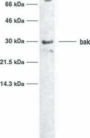
This Anti-Bak (Ab-1) Mouse mAb (TC-100) is validated for use in Immunoblotting for the detection of Bak (Ab-1).
More>> This Anti-Bak (Ab-1) Mouse mAb (TC-100) is validated for use in Immunoblotting for the detection of Bak (Ab-1). Less<<Anti-Bak (Ab-1) Mouse mAb (TC-100) MSDS (material safety data sheet) or SDS, CoA and CoQ, dossiers, brochures and other available documents.
同义词: Anti-Bcl-2 Antagonist Killer
Recommended Products
概述
| Replacement Information |
|---|
重要规格表
| Host | Antibody Type |
|---|---|
| M | Monoclonal Antibody |
价格及供货情况
| 产品目录编号 | 库存情况 | 包装 | 数量 / 包装 | 价格 | 数量 | |
|---|---|---|---|---|---|---|
| AM03-100UGCN |
|
塑胶安瓿;塑胶针药瓶 | 100 μg |
|
— |
| Product Information | |
|---|---|
| Form | Liquid |
| Formulation | In 0.05 M sodium phosphate buffer, 0.2% gelatin. |
| Positive control | HL-60 cells |
| Preservative | ≤0.1% sodium azide |
| Quality Level | MQ200 |
| Physicochemical Information |
|---|
| Dimensions |
|---|
| Materials Information |
|---|
| Toxicological Information |
|---|
| Safety Information according to GHS |
|---|
| Safety Information |
|---|
| Product Usage Statements |
|---|
| Storage and Shipping Information | |
|---|---|
| Ship Code | Blue Ice Only |
| Toxicity | Standard Handling |
| Storage | +2°C to +8°C |
| Do not freeze | Yes |
| Packaging Information |
|---|
| Transport Information |
|---|
| Supplemental Information |
|---|
| Specifications |
|---|
| Global Trade Item Number | |
|---|---|
| 产品目录编号 | GTIN |
| AM03-100UGCN | 04055977227970 |
Documentation
Anti-Bak (Ab-1) Mouse mAb (TC-100) 分析证书
| 标题 | 批号 |
|---|---|
| AM03 |
参考
| 参考信息概述 |
|---|
| Chittenden, T., et al. 1995. Nature 374, 733. Farrow, S.N., et al. 1995. Nature 374, 731. Kiefer, M.C., et al. 1995. Nature 736. Reed, J.C. 1994. J. Cell Biol. 124, 1. Korsmeyer, S.J., et al. 1993. N. Semin. Cancer Biol. 4, 327. |
小册子
| 标题 |
|---|
| Caspases and other Apoptosis Related Tools Brochure |
引用
| 标题 | |
|---|---|
|
|