Millipore Sigma Vibrant Logo
Attention: We have moved. Merck Millipore products are no longer available for purchase on MerckMillipore.com.Learn More

MAB5434-C Anti-Oct-1 Antibody, clone YL15, Ascites Free

View Products on Sigmaaldrich.com
MAB5434-C
100 μg  
Purchase on Sigma-Aldrich

Special Offers

Overview

Replacement Information

Key Spec Table

Species ReactivityKey ApplicationsHostFormatAntibody Type
H, MWB, ICC, IF, IH(P), ChIPMPurifiedMonoclonal Antibody
Description
Catalogue NumberMAB5434-C
ReplacesMAB5434
DescriptionAnti-Oct-1 Antibody, clone YL15, Ascites Free
Alternate Names
  • POU domain, class 2, transcription factor 1
  • NF-A1
  • Oct-1
  • Octamer-binding protein 1
  • Octamer-binding transcription factor 1
  • OTF-1
Background InformationPOU domain, class 2, transcription factor 1 (UniProt P14859; also known as NF-A1, Oct-1, Octamer-binding protein 1, Octamer-binding transcription factor 1, OTF-1) is encoded by the POU2F1 (also known as OCT1, OTF1) gene (Gene ID 5451) in human. Oct-1 belongs to the family of octamer transcription factors characterized by their binding to the ATTTGCAT octamer sequence via their POU domain (a.a. 280-354 of human Oct-1). Oct1 is widely expressed in adult tissues, however, Oct-1 knockdown has little impact on cell growth and viability in culture. Oct-1 controls multiple stem cell phenotypes in normal and tumor cells. Oct-1 promotes resistance to genotoxic and oxidative stresses in stem cells. Loss of Oct-1 antagonizes oncogenic transformation in vitro and tumorigenicity in vivo.
References
Product Information
FormatPurified
PresentationPurified mouse monoclonal IgG1κ antibody in buffer containing 0.1 M Tris-Glycine (pH 7.4), 150 mM NaCl with 0.05% sodium azide.
Quality LevelMQ100
Applications
ApplicationAnti-Oct-1 Antibody, clone YL15, Cat. No. MAB5434-C, is a mouse monoclonal antibody that detects octamer-binding protein 1 (Oct-1) and has been tested for use in immunocytochemistry, immunohistchemistry (Parffin), Immunoflouroesencece, Western blotting, and Chromatin Immunorecipitation.
Key Applications
  • Western Blotting
  • Immunocytochemistry
  • Immunofluorescence
  • Immunohistochemistry (Paraffin)
  • Chromatin Immunoprecipitation (ChIP)
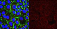
Application NotesImmunocytochemistry Analysis: 5.0 µg/mL from a representative lot detected Oct-1 cellular localization in human A431 and HeLa cells by fluorescent immunocytochemistry.
Western Blotting Analysis: A representative lot detected Oct-1, but not Oct-2, Oct-4, or Oct-6 in various human cell lysates and nuclear extracts (Maddox, J., et al. (2012). PLoS Genet. 8(11):e1003048).
Western Blotting Analysis: A representative lot detected Oct-1 in Burkitt lymphoma cells (BJAB and Namalwa), as well as a panel of six primary effusion lymphoma (PEL) cells (Di Bartolo, D.L., et al. (2009). J. Virol. 83(9):4308-4315).
Immunofluorescence Analysis: A representative lot detected Oct-1 immunoreactivity in paraformaldehyde-fixed human and mouse frozen colon tissue sections by fluorescent immunohistochemistry (Maddox, J., et al. (2012). PLoS Genet. 8(11):e1003048).
Immunohistochemistry Analysis: A representative lot detected Oct-1 immunoreactivity in formalin-fixed, paraffin-embedded human colon tissue sections (Maddox, J., et al. (2012). PLoS Genet. 8(11):e1003048).
Chromatin Immunoprecipitation (ChIP) Analysis: A representative lot detected Oct-1 occupancy at the ORF50 promoter, using stable Oct-2-inducible primary effusion lymphoma (PEL) cell line BC1/Tet-on/Oct-2. The association decreased upon induction of Oct-2 expression and enhanced upon ORF50 transfection (Di Bartolo, D.L., et al. (2009). J. Virol. 83(9):4308-4315).
Chromatin Immunoprecipitation (ChIP) Analysis: A representative lot detected Oct-1 occupancy at the U6 and U1 promoter regions in exponentially dividing HeLa cells (Zhao, X., et al. (2001). Mol. Cell. 7(3):539-549).
Electrophoretic Mobility Shift Assay (EMSA): A representative lot caused a supershift of radiolabeled OCT1 consensus oligonucleotide-bound complex in the nuclear extracts from Burkitt lymphoma cells BJAB and Namalwa (Di Bartolo, D.L., et al. (2009). J. Virol. 83(9):4308-4315).
Immunocytochemistry Analysis: A representative lot detected Oct-1 cellular localization in human diploid fibroblast MRC-5-derived HuS-L12 cells at various population-doubling levels (PDLs), as well as in the corresponding immortalized IML12–4 cells by fluorescent immunocytochemistry (Imai, S., et al. (1997). Mol. Biol. Cell. 8(12):2407-2419).
Immunoprecipitation Analysis: A representative lot immunoprecipitated Oct-1 from HEK293 cell lysates (Lai, J.S., and Herr, W. (1992). Proc. Natl. Acad. Sci. U. S. A. 89(15):6958-6962).
Biological Information
ImmunogenRecombinant full-length human Oct-1.
EpitopePOU-domain linker region.
CloneYL15
ConcentrationPlease refer to lot specific datasheet.
HostMouse
SpecificityClone YL15 targets the POU-domain linker region (Herr, W., and Cleary, M.A. (1995). Genes Dev. 9(14):1679-1693; Lai, J.S., and Herr, W. (1992). Proc. Natl. Acad. Sci. U. S. A. 89(15):6958-6962) present in all six spliced isoforms of human Oct-1 (UniProt P14859). Clone YL15 does not react with Oct-2, Oct-4 or Oct-6 (Maddox, J., et al. (2012). PLoS Genet. 8(11):e1003048).
IsotypeIgG1κ
Species Reactivity
  • Human
  • Mouse
Antibody TypeMonoclonal Antibody
Entrez Gene Number
Gene Symbol
  • POU2F1
  • OCT1
  • OTF1
Purification MethodProtein G Purified
UniProt Number
Molecular Weight~96 kDa observed.
Physicochemical Information
Dimensions
Materials Information
Toxicological Information
Safety Information according to GHS
Safety Information
Product Usage Statements
Quality AssuranceEvaluated by Western Blotting in HeLa cell lysate.

Western Blotting Analysis: 2.0 µg/mL of this antibody detected Oct-1 in 10 µg of HeLa cell lysate.
Usage Statement
  • Unless otherwise stated in our catalog or other company documentation accompanying the product(s), our products are intended for research use only and are not to be used for any other purpose, which includes but is not limited to, unauthorized commercial uses, in vitro diagnostic uses, ex vivo or in vivo therapeutic uses or any type of consumption or application to humans or animals.
Storage and Shipping Information
Storage ConditionsStable for 1 year at 2-8°C from date of receipt.
Packaging Information
Material Size100 μg
Transport Information
Supplemental Information
Specifications
Global Trade Item Number
Catalogue Number GTIN
MAB5434-C 04055977333206