Le fait de fermer ne sauvegardera pas votre configuration à moins que vous n'ajoutiez l'article à votre Panier d'achat ou à vos Favoris.
Cliquer sur OK pour fermer l'outil MILLIPLEX® MAP ou sur Annuler pour retourner à votre sélection.
Choisissez des Panels configurables & des Kits préconfigurés - OU - des MAPmate™ de signalisation cellulaire
Concevez vos kits MILLIPLEX® MAP et obtenez leur prix.
Panels configurables & Kits préconfigurés
Notre large gamme est constituée de panels multiplex qui vous permettent de choisir, au sein d'un panel, les analytes qui répondent le mieux à vos besoins. Sur un autre onglet, vous pouvez choisir un format cytokine préconfiguré ou un kit Simplex.
Kits de signalisation cellulaire & MAPmate™
Choisissez des kits préconfigurés qui permettent d'explorer l'ensemble des voies ou des processus. Ou concevez vos propres kits en choisissant des Simplex MAPmate™ et en suivant les instructions fournies.
Les MAPmate™ suivants ne peuvent pas être utilisés ensemble : -des MAPmate™ qui nécessitent des tampons différents -des paires de MAPmate™ totaux et phospho-spécifiques, par ex. GSK3β total et GSK3β (Ser 9) -des MAPmate™ PanTyr et spécifiques d'un site, par ex. Récepteur Phospho-EGF et phospho-STAT1 (Tyr701) -Plus d'un phospho-MAPmate™ pour une seule cible (Akt, STAT3). -GAPDH et β-Tubuline ne peuvent pas être utilisés avec les kits ou les MAPmate™ contenant panTyr.
.
Référence
Guide d'achat
Qté
Liste
Cet article a été ajouté à vos favoris.
Sélectionner une espèce, un type de panel, un kit ou un type d'échantillon
Pour commencer à concevoir votre kit MILLIPLEX® MAP, sélectionnez une espèce, un type de panel ou un kit d'intérêt.
Custom Premix Selecting "Custom Premix" option means that all of the beads you have chosen will be premixed in manufacturing before the kit is sent to you.
Catalogue Number
Ordering Description
Qty/Pack
List
Cet article a été ajouté à vos favoris.
Espèce
Type de panel
Kit sélectionné
Qté
Référence
Guide d'achat
Qté
Prix tarif
96-Well Plate
Qté
Référence
Guide d'achat
Qté
Prix tarif
Ajouter des réactifs supplémentaires (Un kit "Buffer and Detection Kit" est nécessaire pour une utilisation avec les MAPmate™)
Qté
Référence
Guide d'achat
Qté
Prix tarif
48-602MAG
Buffer Detection Kit for Magnetic Beads
1 Kit
Option de gain de place Nos clients qui commandent plusieurs kits peuvent choisir d'économiser de l'espace de stockage en éliminant l'emballage de chaque kit et de recevoir les composants de leur essai multiplex conditionnés sous poches en plastique pour un stockage plus compact.
Cet article a été ajouté à vos favoris.
Ce produit a été ajouté à votre panier.
Vous pouvez maintenant concevoir un autre kit personnalisé, choisir un kit pré-configuré, régler vos achats ou fermer l'outil de commande.
ST1029
Sigma-AldrichPhosphoDetect™ Anti-Fos (pSer³⁷⁴) Mouse mAb (34E4)
This PhosphoDetect™ Anti-Fos (pSer³⁷⁴) Mouse mAb (34E4) is validated for use in ELISA, Immunoblotting for the detection of Fos (pSer³⁷⁴).
More>>This PhosphoDetect™ Anti-Fos (pSer³⁷⁴) Mouse mAb (34E4) is validated for use in ELISA, Immunoblotting for the detection of Fos (pSer³⁷⁴). Less<<
FDS (Fiches de données de sécurité), certificats d’analyse (CoA) et de qualité (CoQ), dossiers, brochures et autres documents disponibles.
Le champ quantité est vide. Veuillez saisir une quantité supérieure ou égale à 1 au minimum pour ajouter des articles à votre panier.
Description
Overview
Recognizes the ~55 kDa c-Fos protein phosphorylated at Ser374. Does not detect the unphosphorylated form.
Catalogue Number
ST1029
Brand Family
Calbiochem®
Application Data
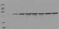
Detection of human Fos, phosphorylated on Ser374, by immunoblotting. Samples: Serum starved HepG2 cells incubated with EGF for 0 min (lane 1), 5 min (lane 2), 15 min (lane 3), 30 min (lane 4), 1 h (lane 5), 2 h (lane 6), 4 h (lane 7), and 8 h (lane 8). Primary antibody: PhosphoDetect™ Anti-Fos (pSer374) Mouse mAb (34E4) (Cat. No. ST1029) (0.5 µg/ml). Detection: chemiluminescence.
References
References
Chen, R-H., et al. 1993. Proc. Natl. Acad. Sci. USA90, 10952.
Product Information
Form
Lyophilized
Formulation
100 µg antibody lyophilized from 2X PBS, PEG, sucrose and 200 µl lyophilized control lysate from EGF-treated HEPG2 cells.
Does not detect the unphosphorylated form of c-fos. For immunoblotting use 20 µl control lysate per lane (minigel) for chemiluminescent detection. Antibody should be titrated for optimal results in individual systems.
Biological Information
Immunogen
a synthetic phosphopeptide corresponding to amino acids surrounding the Ser³⁷⁴ phosphorylation site of human c-Fos
Immunogen
Human
Clone
34E4
Host
Mouse
Isotype
IgG₁
Species Reactivity
Human
Antibody Type
Monoclonal Antibody
Physicochemical Information
Dimensions
Materials Information
Toxicological Information
Safety Information according to GHS
Safety Information
Product Usage Statements
Storage and Shipping Information
Ship Code
Shipped with Blue Ice or with Dry Ice
Toxicity
Irritant
Storage
-20°C
Avoid freeze/thaw
Avoid freeze/thaw
Do not freeze
Ok to freeze
Special Instructions
Reconstitute antibody with 1 ml water (15 minutes, room temperature). Following reconstitution, aliquot and freeze in liquid nitrogen. Reconstituted antibody can be stored at -80°C for up to 1 year. Aliquots may be stored at 4°C for up to 3 months and should be thawed at 37°C. Reconstitute control lysate with 200 µl H₂O. After complete solubilization of the proteins add 200 µl SDS-PAGE sample buffer and incubate at 90°C for 5 min. Following reconsitution aliquot and freeze (-20°C). Avoid freeze/thaw cycles.
Packaging Information
Transport Information
Supplemental Information
Specifications
Global Trade Item Number
Référence
GTIN
ST1029-1SET
04055977208689
Documentation
PhosphoDetect™ Anti-Fos (pSer³⁷⁴) Mouse mAb (34E4) FDS
PhosphoDetect™ Anti-Fos (pSer³⁷⁴) Mouse mAb (34E4) Certificats d'analyse
Titre
Numéro de lot
ST1029
Références bibliographiques
Aperçu de la référence bibliographique
Chen, R-H., et al. 1993. Proc. Natl. Acad. Sci. USA90, 10952.
Fiche technique
Note that this data sheet is not lot-specific and is representative of the current specifications for this product. Please consult the vial label and the certificate of analysis for information on specific lots. Also note that shipping conditions may differ from storage conditions.
Detection of human Fos, phosphorylated on Ser374, by immunoblotting. Samples: Serum starved HepG2 cells incubated with EGF for 0 min (lane 1), 5 min (lane 2), 15 min (lane 3), 30 min (lane 4), 1 h (lane 5), 2 h (lane 6), 4 h (lane 7), and 8 h (lane 8). Primary antibody: PhosphoDetect™ Anti-Fos (pSer374) Mouse mAb (34E4) (Cat. No. ST1029) (0.5 µg/ml). Detection: chemiluminescence.
Description
Mouse monoclonal antibody purified from serum-free cell culture supernatant by thiophilic adsorption and size exclusion chromatography. Supplied with a positive control lysate consisting of EGF-treated HepG2 cells. Recognizes the ~55 kDa c-fos protein phosphorylated at Ser374 by MAP kinase.
Background
The early gene product c-Fos is expressed following mitogenic stimulation and functions as a sensor for MAPK signal duration. When MAPK activation is transient, MAPK activity declines before accumulation of the c-Fos protein. When MAPK activation is sustained, c-Fos is phosphorylated by MAPK at Ser374. Phosphorylation stabilizes the Fos protein and primes c-Fos for additional phosphorylation at Thr325.
Host
Mouse
Immunogen species
Human
Immunogen
a synthetic phosphopeptide corresponding to amino acids surrounding the Ser³⁷⁴ phosphorylation site of human c-Fos
Clone
34E4
Isotype
IgG₁
Species
human
Positive control
HepG2 cells treated with EGF
Form
Lyophilized
Formulation
100 µg antibody lyophilized from 2X PBS, PEG, sucrose and 200 µl lyophilized control lysate from EGF-treated HEPG2 cells.
Preservative
≤0.1% sodium azide (antibody only)
Comments
Does not detect the unphosphorylated form of c-fos. For immunoblotting use 20 µl control lysate per lane (minigel) for chemiluminescent detection. Antibody should be titrated for optimal results in individual systems.
Storage
Avoid freeze/thaw
-20°C
Do Not Freeze
Ok to freeze
Special Instructions
Reconstitute antibody with 1 ml water (15 minutes, room temperature). Following reconstitution, aliquot and freeze in liquid nitrogen. Reconstituted antibody can be stored at -80°C for up to 1 year. Aliquots may be stored at 4°C for up to 3 months and should be thawed at 37°C. Reconstitute control lysate with 200 µl H₂O. After complete solubilization of the proteins add 200 µl SDS-PAGE sample buffer and incubate at 90°C for 5 min. Following reconsitution aliquot and freeze (-20°C). Avoid freeze/thaw cycles.
Toxicity
Irritant
References
Chen, R-H., et al. 1993. Proc. Natl. Acad. Sci. USA90, 10952.