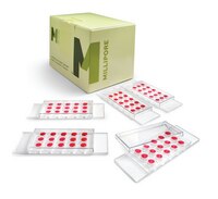
MMA125 MilliporeMillicell® µ-Angiogenesis Inhibition Assay Kit
The Millicell μ-Angiogenesis inhibition assay kits provide a quantitative platform for real-time monitoring of changes in tubule formation with unprecedented optical resolution.
More>> The Millicell μ-Angiogenesis inhibition assay kits provide a quantitative platform for real-time monitoring of changes in tubule formation with unprecedented optical resolution. Less<<Produits recommandés
Aperçu
| Replacement Information |
|---|
Tableau de caractéristiques principal
| Key Applications | Detection Methods |
|---|---|
| ACT, CULT, INHIB | Fluorescent |
| Description | |
|---|---|
| Catalogue Number | MMA125 |
| Trade Name |
|
| Description | Millicell® µ-Angiogenesis Inhibition Assay Kit |
| Overview | Overview Studying how compounds affect angiogenesis, either to promote or inhibit new capillary tube formation can lead to therapies affecting wound healing, tissue regeneration, cardiovascular disease, stroke, tumor progression, and more. The Millicell μ-Angiogenesis activation and inhibition kits provide a powerful, quantitative platform for real-time monitoring of changes in tubule formation with unprecedented optical resolution. Benefits
Studying how compounds affect angiogenesis, either to promote or inhibit new capillary tube formation, can lead to therapies affecting wound healing, tissue regeneration, cardiovascular disease, stroke, tumor progression, and more. The Millicell® μ-Angiogenesis activation and inhibition assay kits provide a powerful, quantitative platform for real-time monitoring of changes in tubule formation with unprecedented optical resolution. Applications
For more information on Cell Culture & Systems visit: www.emdmillipore.com/cellculture |
| Materials Required but Not Delivered | 1. Inverted Light Microscope 2. Fluorescence microscope (if Calcein AM is used) 3. Precision pipettes 4. Accutase™ Cell Dissociation Solution (Catalog. No. SCR005) 5. Human umbilical vein endothelial cells (HUVEC) (Catalog. No. SCCE001) or any other experimental cell lines capable of tube formation 6. EndoGRO™ LS complete medium (Catalog. No. SCME001) or other endothelial cell basal medium. 7. Phosphate-Buffered Saline (1X PBS) (Catalog No. BSS-1005-B) 8. EmbryoMax ES Cell Qualified Ultra Pure Water, sterile H20, 500 mL (Catalog No. TMS-006-B) 9. Low speed centrifuge 10. Sterile eppendorf tubes 11. 37ºC Incubator with 5% CO2 12. Hemacytometer 13. Trypan Blue 14. DMSO |
| Background Information | Angiogenesis, the formation of new blood vessels from a pre-existing vascular network, occurs normally in development and is critical for a majority of the vessel formation that occurs during embryogenesis, tissue generation, and wound healing. However, abnormal blood vessel growth, either excessive or insufficient, can be the underlying cause of many deadly and debilitating diseases including cancer, cardiovascular disease, stroke, and diabetic and age-related blindness. Identification of specific compounds that promote or inhibit angiogenesis may provide promising treatments for these diseases. Tubular formation is a multi-step process involving cell adhesion, migration, differentiation, and growth. Millipore’s Millicell® µ-Angiogenesis Inhibition Assay provides an efficient system for the rapid and accurate identification of factors that inhibit tube formation of endothelial cells. The µ-Angiogenesis slide, with enhanced optical capabilities in a microscale format, allows for real time and continuous monitoring of tube formation in a simplifed and reproducible manner. |
| References |
|---|
| Biological Information | |
|---|---|
| Species Reactivity |
|
| Physicochemical Information |
|---|
| Dimensions |
|---|
| Materials Information |
|---|
| Toxicological Information |
|---|
| Safety Information according to GHS |
|---|
| Safety Information |
|---|
| Packaging Information | |
|---|---|
| Material Size | 1 Kit |
| Transport Information |
|---|
| Supplemental Information |
|---|
| Specifications |
|---|
| Global Trade Item Number | |
|---|---|
| Référence | GTIN |
| MMA125 | 04053252006333 |
Documentation
Brochure
| Titre |
|---|
| EpiGRO™, EndoGRO™ and FibroGRO™ Reagents |
Posters
| Titre |
|---|
| Poster: Tumor Metastasis |
Manuels d'utilisation
| Titre |
|---|
| Millicell -Angiogenesis Inhibition Assay |


