KSRP modulation of GAP-43 mRNA stability restricts axonal outgrowth in embryonic hippocampal neurons.
Bird, CW; Gardiner, AS; Bolognani, F; Tanner, DC; Chen, CY; Lin, WJ; Yoo, S; Twiss, JL; Perrone-Bizzozero, N
PloS one
8
e79255
2013
Show Abstract
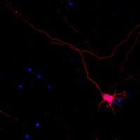
The KH-type splicing regulatory protein (KSRP) promotes the decay of AU-rich element (ARE)-containing mRNAs. Although KSRP is expressed in the nervous system, very little is known about its role in neurons. In this study, we examined whether KSRP regulates the stability of the ARE-containing GAP-43 mRNA. We found that KSRP destabilizes this mRNA by binding to its ARE, a process that requires the presence of its fourth KH domain (KH4). Furthermore, KSRP competed with the stabilizing factor HuD for binding to these sequences. We also examined the functional consequences of KSRP overexpression and knockdown on the differentiation of primary hippocampal neurons in culture. Overexpression of full length KSRP or KSRP without its nuclear localization signal hindered axonal outgrowth in these cultures, while overexpression of a mutant protein without the KH4 domain that has less affinity for binding to GAP-43's ARE had no effect. In contrast, depletion of KSRP led to a rise in GAP-43 mRNA levels and a dramatic increase in axonal length, both in KSRP shRNA transfected cells and neurons cultured from Ksrp(+/-) and Ksrp(-/-) embryos. Finally we found that overexpression of GAP-43 rescued the axonal outgrowth deficits seen with KSRP overexpression, but only when cells were transfected with GAP-43 constructs containing 3' UTR sequences targeting the transport of this mRNA to axons. Together, our results suggest that KSRP is an important regulator of mRNA stability and axonal length that works in direct opposition to HuD to regulate the levels of GAP-43 and other ARE-containing neuronal mRNAs. | | 24244461
 |
A ROCK Inhibitor Blocks the Inhibitory Effect of Chondroitin Sulfate Proteoglycan on Morphological Changes of Mesenchymal Stromal/Stem Cells into Neuron-Like Cells.
Lim, HS; Joe, YA
Biomolecules & therapeutics
21
447-53
2013
Show Abstract
Chondroitin sulfate proteoglycan (CSPG) inhibits neurite outgrowth of various neuronal cell types, and CSPG-associated inhibition of neurite outgrowth is mediated by the Rho/ROCK pathway. Mesenchymal stromal/stem cells (MSCs) have the potential to differentiate into neuron-like cells under specific conditions and have been shown to differentiate into neuron-like cells by co-treatment with the ROCK inhibitor Y27632 and the hypoxia condition mimicking agent CoCl2. In this study, we addressed the hypothesis that a ROCK inhibitor might be beneficial to regenerate neurons during stem cell therapy by preventing transplanted MSCs from inhibition by CSPG in damaged tissues. Indeed, dose-dependent inhibition by CSPG pretreatment was observed during morphological changes of Wharton's jelly-derived MSCs (WJ-MSCs) induced by Y27632 alone. The formation of neurite-like structures was significantly inhibited when WJ-MSCs were pre-treated with CSPG before induction under Y27632 plus CoCl2 conditions, and pretreatment with a protein kinase C inhibitor reversed such inhibition. However, CSPG treatment resulted in no significant inhibition of the WJ-MSC morphological changes into neuron-like cells after initiating induction by Y27632 plus CoCl2. No marked changes were detected in expression levels of neuronal markers induced by Y27632 plus CoCl2 upon CSPG treatment. CSPG also blocked the morphological changes of human bone marrow-derived MSCs into neuron-like cells under other neuronal induction condition without the ROCK inhibitor, and Y27632 pre-treatment blocked the inhibitory effect of CSPG. These results suggest that a ROCK inhibitor can be efficiently used in stem cell therapy for neuronal induction by avoiding hindrance from CSPG. | | 24404335
|
Primed pluripotent cell lines derived from various embryonic origins and somatic cells in pig.
Park, JK; Kim, HS; Uh, KJ; Choi, KH; Kim, HM; Lee, T; Yang, BC; Kim, HJ; Ka, HH; Kim, H; Lee, CK
PloS one
8
e52481
2013
Show Abstract
Since pluripotent embryonic stem cell (ESC) lines were first derived from the mouse, tremendous efforts have been made to establish ESC lines in several domestic species including the pig; however, authentic porcine ESCs have not yet been established. It has proven difficult to maintain an ESC-like state in pluripotent porcine cell lines due to the frequent occurrence of spontaneous differentiation into an epiblast stem cell (EpiSC)-like state during culture. We have been able to derive EpiSC-like porcine ESC (pESC) lines from blastocyst stage porcine embryos of various origins, including in vitro fertilized (IVF), in vivo derived, IVF aggregated, and parthenogenetic embryos. In addition, we have generated induced pluripotent stem cells (piPSCs) via plasmid transfection of reprogramming factors (Oct4, Sox2, Klf4, and c-Myc) into porcine fibroblast cells. In this study, we analyzed characteristics such as marker expression, pluripotency and the X chromosome inactivation status in female of our EpiSC-like pESC lines along with our piPSC line. Our results show that these cell lines demonstrate the expression of genes associated with the Activin/Nodal and FGF2 pathways along with the expression of pluripotent markers Oct4, Sox2, Nanog, SSEA4, TRA 1-60 and TRA 1-81. Furthermore all of these cell lines showed in vitro differentiation potential, the X chromosome inactivation in female and a normal karyotype. Here we suggest that the porcine species undergoes reprogramming into a primed state during the establishment of pluripotent stem cell lines. | | 23326334
|
TrkB signaling directs the incorporation of newly generated periglomerular cells in the adult olfactory bulb.
Bergami, M; Vignoli, B; Motori, E; Pifferi, S; Zuccaro, E; Menini, A; Canossa, M
The Journal of neuroscience : the official journal of the Society for Neuroscience
33
11464-78
2013
Show Abstract
In the adult rodent brain, the olfactory bulb (OB) is continuously supplied with new neurons which survival critically depends on their successful integration into pre-existing networks. Yet, the extracellular signals that determine the selection which neurons will be ultimately incorporated into these circuits are largely unknown. Here, we show that immature neurons express the catalytic form of the brain-derived neurotrophic factor receptor TrkB [full-length TrkB (TrkB-FL)] only after their arrival in the OB, at the time when integration commences. To unravel the role of TrkB signaling in newborn neurons, we conditionally ablated TrkB-FL in mice via Cre expression in adult neural stem and progenitor cells. TrkB-deficient neurons displayed a marked impairment in dendritic arborization and spine growth. By selectively manipulating the signaling pathways initiated by TrkB in vivo, we identified the transducers Shc/PI3K to be required for dendritic growth, whereas the activation of phospholipase C-γ was found to be responsible for spine formation. Furthermore, long-term genetic fate mapping revealed that TrkB deletion severely compromised the survival of new dopaminergic neurons, leading to a substantial reduction in the overall number of adult-generated periglomerular cells (PGCs), but not of granule cells (GCs). Surprisingly, this loss of dopaminergic PGCs was mirrored by a corresponding increase in the number of calretinin+ PGCs, suggesting that distinct subsets of adult-born PGCs may respond differentially to common extracellular signals. Thus, our results identify TrkB signaling to be essential for balancing the incorporation of defined classes of adult-born PGCs and not GCs, reflecting their different mode of integration in the OB. | | 23843518
|
Brilliant violet fluorophores: a new class of ultrabright fluorescent compounds for immunofluorescence experiments.
Pratip K Chattopadhyay,Brent Gaylord,Adrian Palmer,Nan Jiang,Mary A Raven,Geoff Lewis,Morgan A Reuter,A K M Nur-Ur Rahman,David A Price,Michael R Betts,Mario Roederer
Cytometry. Part A : the journal of the International Society for Analytical Cytology
81
2012
Show Abstract
The Nobel Prize in Chemistry was awarded in 2000 for the discovery of conductive organic polymers, which have subsequently been adapted for applications in ultrasensitive biological detection. Here, we report the first use of this new class of fluorescent probes in a diverse range of cytometric and imaging applications. We demonstrate that these Brilliant Violet reporters are dramatically brighter than other UV-violet excitable dyes, and are of similar utility to phycoerythrin (PE) and allophycocyanin (APC). They are thus ideally suited for cytometric assays requiring high sensitivity, such as MHC-multimer staining or detection of intracellular antigens. Furthermore, these reporters are sensitive and spectrally distinct options for fluorescence imaging, two-photon microscopy and imaging cytometry. These ultra-bright materials provide the first new high-sensitivity fluorescence probes in over 25 years and will have a dramatic impact on the design and implementation of multicolor panels for high-sensitivity immunofluorescence assays. | | 22489009
|
Investigation of cytoskeleton proteins in neurons of the cat lateral geniculate nucleus.
Duffy KR, Crowder NA, Ledue EE
The Journal of comparative neurology
520
186-99. doi
2012
Show Abstract
The gross structure of neurons is supported by proteins that compose the cytoskeleton. Neurofilaments are intermediate cytoskeletal proteins that contribute to neuron structure and function, and three neurofilament subunits different in their molecular mass assemble to form heteropolymers that produce a structure-providing intracellular scaffold. The light neurofilament subunit is obligatory and can assemble with either the medium or heavy subunit, indicating some degree of independence between subunits. The presence of the heavy subunit has been shown to be associated with mature cells and is linked to large neurons in the cerebral cortex and thalamus. Spectrin is a membrane-associated actin-binding protein that, like neurofilament, has been linked to neuron shape. In this study of the cat dorsal lateral geniculate nucleus (dLGN) we examined whether labeling for neurofilament subunits and spectrin is linked to neuron size. We found that about one-third of neurons contained a visible amount of labeling for each neurofilament subunit, and the bulk of these labeled cells were large in comparison to the general population of neurons. The distribution of neuron sizes was not different between neurofilament subunits, indicating that neurofilament subunit content is not determined by neuron size. Spectrin labeling was evident in most dLGN neurons, and was not related to the size of neurons. That reactivity for neurofilament was predominant in large cells led us to directly examine the relationship between neurofilament and interneurons. The large majority of neurofilament-positive neurons did not contain GABA, indicating that neurofilament is predominant in projection cells and not in interneurons. J. Comp. Neurol., 2012. © 2011 Wiley Periodicals, Inc.Copyright © 2011 Wiley Periodicals, Inc. | | 21800310
|
Beneficial effects of exendin-4 on experimental polyneuropathy in diabetic mice.
Himeno, T; Kamiya, H; Naruse, K; Harada, N; Ozaki, N; Seino, Y; Shibata, T; Kondo, M; Kato, J; Okawa, T; Fukami, A; Hamada, Y; Inagaki, N; Seino, Y; Drucker, DJ; Oiso, Y; Nakamura, J
Diabetes
60
2397-406
2011
Show Abstract
The therapeutic potential of exendin-4, an agonist of the glucagon-like peptide-1 receptor (GLP-1R), on diabetic polyneuropathy (DPN) in streptozotocin (STZ)-induced diabetic mice was investigated.The presence of the GLP-1R in lumbar dorsal root ganglion (DRG) was evaluated by immunohistochemical analyses. DRG neurons were dissected from C57BL6/J mice and cultured with or without Schwann cell-conditioned media in the presence or absence of GLP-1 (7-37) or exendin-4. Then neurite outgrowth was determined. In animal-model experiments, mice were made diabetic by STZ administration, and after 12 weeks of diabetes, exendin-4 (10 nmol/kg) was intraperitoneally administered once daily for 4 weeks. Peripheral nerve function was determined by the current perception threshold and motor and sensory nerve conduction velocity (MNCV and SNCV, respectively). Sciatic nerve blood flow (SNBF) and intraepidermal nerve fiber densities (IENFDs) also were evaluated.The expression of the GLP-1R in DRG neurons was confirmed. GLP-1 (7-37) and exendin-4 significantly promoted neurite outgrowth of DRG neurons. Both GLP-1R agonists accelerated the impaired neurite outgrowth of DRG neurons cultured with Schwann cell-conditioned media that mimicked the diabetic condition. At the doses used, exendin-4 had no effect on blood glucose or HbA(1c) levels. Hypoalgesia and delayed MNCV and SNCV in diabetic mice were improved by exendin-4 without affecting the reduced SNBF. The decreased IENFDs in sole skins of diabetic mice were ameliorated by exendin-4.Our findings indicate that exendin-4 ameliorates the severity of DPN, which may be achieved by its direct actions on DRG neurons and their axons. | | 21810596
|
Cytoarchitecture of the mouse neocortex revealed by the low-molecular-weight neurofilament protein subunit.
Melissa Paulussen,Sandy Jacobs,Estelle Van der Gucht,Patrick R Hof,Lutgarde Arckens
Brain structure & function
216
2011
Show Abstract
The expression patterns of the medium- and high-molecular-weight subunits of the neurofilament protein triplet have been extensively studied in several neuroanatomical studies. In the present study, we report the use of the low-molecular-weight neurofilament protein subunit (NF-L) as a reliable marker within the neurofilament protein family to reveal the regional architecture of mammalian neocortex. We document clearly its usefulness in anatomical parcellation studies and report unique expression patterns of NF-L throughout the mouse neocortex. NF-L was most abundant in the somatosensory cortex, the lateral secondary visual area, the granular insular cortex, and the motor cortex. Low NF-L staining intensity was observed in the agranular insular cortex, the prelimbic and infralimbic cortex, the anterior cingulate cortex, the visual rostromedial areas, the temporal association cortex, the ectorhinal cortex, and the lateral entorhinal cortex. NF-L immunoreactivity was present in the perikarya, dendrites, and proximal segment of axons primarily of pyramidal neurons, and was mainly located in layers II and III, and to a lesser extent in layers V and VI. Interestingly, Black-Gold myelin staining confirmed a close correlation between NF-L immunoreactivity and myelination patterns. The characteristic and distinctive distribution and laminar expression profiles of NF-L make it an excellent tool to assess accurately topographical boundaries among neocortical areas as illustrated herein in the adult mouse brain. | | 21465412
|
Differentiation of induced pluripotent stem cells into functional oligodendrocytes.
Czepiel M, Balasubramaniyan V, Schaafsma W, Stancic M, Mikkers H, Huisman C, Boddeke E, Copray S
Glia
59
882-92
2011
Show Abstract
The technology to generate autologous pluripotent stem cells (iPS cells) from almost any somatic cell type has brought various cell replacement therapies within clinical research. Besides the challenge to optimize iPS protocols to appropriate safety and GMP levels, procedures need to be developed to differentiate iPS cells into specific fully differentiated and functional cell types for implantation purposes. In this article, we describe a protocol to differentiate mouse iPS cells into oligodendrocytes with the aim to investigate the feasibility of IPS stem cell-based therapy for demyelinating disorders, such as multiple sclerosis. Our protocol results in the generation of oligodendrocyte precursor cells (OPCs) that can develop into mature, myelinating oligodendrocytes in-vitro (co-culture with DRG neurons) as well as in-vivo (after implantation in the demyelinated corpus callosum of cuprizone-treated mice). We report the importance of complete purification of the iPS-derived OPC suspension to prevent the contamination with teratoma-forming iPS cells. © 2011 Wiley-Liss, Inc. Copyright © 2011 Wiley-Liss, Inc. | | 21438010
|
Progesterone potentiates calcium release through IP3 receptors by an Akt-mediated mechanism in hippocampal neurons.
Hwang, JY; Duncan, RS; Madry, C; Singh, M; Koulen, P
Cell calcium
45
233-42
2009
Show Abstract
Progesterone (P4) is a steroid hormone that plays multiple roles in the central nervous system (CNS) including promoting neuroprotection. However, the precise mechanisms involved in its neuroprotective effects are still unknown. Given that the regulation of the intracellular calcium (Ca(2+)) concentration is critical for cell survival, we determined if inositol 1, 4, 5-trisphosphate receptors (IP(3)Rs) are relevant targets of P4. Using primary hippocampal neurons, we tested the hypothesis that P4 controls the gain of IP3R-mediated intracellular Ca(2+) signaling in neurons and characterized the subcellular distribution and phosphorylation of potential signaling intermediates involved in P4s actions. Our results reveal that P4 treatment altered the intensity and distribution of IP3R immunoreactivity and induced the nuclear translocation of phosphorylated Akt. Further, P4 potentiated IP(3)R-mediated intracellular Ca(2+) responses. These results suggest a potential involvement of P4 in particular and of steroid hormone signaling pathways in general in the control of intracellular Ca(2+) signaling and its related functions. Full Text Article | | 19081133
|