Millipore Sigma Vibrant Logo
Atención: Nos hemos mudado. Los productos Merck Millipore ya no pueden adquirirse en MerckMillipore.comMás información

506123 Anti-p38 MAP Kinase (341-360) Rabbit pAb

506123
Purchase on Sigma-Aldrich

Descripción

Replacement Information

Tabla espec. clave

Species ReactivityHostAntibody Type
H, M, RRbPolyclonal Antibody

Products

Número de referenciaEmbalaje Cant./Env.
506123-200UL Ampolla de plást. 200 ul
Description
OverviewRecognizes the ~38 kDa p38 MAPK protein.
  • Antibody Target Gene Symbol: MAPK14
  • Target Synonym: CRK1, CSBP, CSBP1, CSBP2, CSPB1, EXIP, Hog, MAPK p38, MGC102436, MGC105413, MXI2, P38, P38 KINASE, P38 Map Kinase, p38 Mapk alpha, P38-ALPHA, p38-RK, p38/Hog1, p38/Mpk2, P38/RK, p38a, p38Hog, p38MAPK, PRKM14, PRKM15, RK, SAPK2A
  • Entrez Gene Name: mitogen-activated protein kinase 14
  • Hu Entrez ID: 1432
  • Mu Entrez ID: 26416
  • Rat Entrez ID: 81649
  • Catalogue Number506123
    Brand Family Calbiochem®
    Application Data
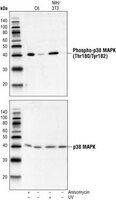
    Detection of p38 MAP kinase by immunoblotting. Samples: C6 cells (lanes 1 and 2) treated (lane 1) or untreated (lane 2) with anisomycin and NIH/3T3 cells (lanes 3 and 4) treated (lane 3) or untreated (lane 4) with UV. Primary antibody: Anti-p38 MAP Kinase (341-360) Rabbit pAb (Cat. No. 506123) (1:1000). Detection: chemiluminescence.
    References
    ReferencesRaingeaud, J., et al. 1995. J. Biol. Chem. 270, 7420.
    Zervos, A.S., et al. 1995. Proc. Natl. Acad. Sci. USA 92, 10531.
    Han, J., et al. 1994. Science 265, 808.
    Lee, J.C., et al. 1994. Nature 372, 739.
    Rouse, J., et al. 1994. Cell 78, 1027.
    Product Information
    FormLiquid
    FormulationIn 150 mM NaCl, 10 mM HEPES, 50% glycerol, 0.01% BSA, pH 7.5.
    PreservativeNone
    Quality LevelMQ100
    Applications
    Key Applications Flow Cytometry
    Immunoblotting (Western Blotting)
    Paraffin Sections
    Application NotesFlow Cytometry (1:25)
    Immunoblotting (1:1000)
    Paraffin Sections (1:50, heat pretreatment required, see comments)
    Application CommentsPretreat paraffin sections by heating tissue in 10 mM citrate buffer, pH 6.0 for 1 min at high power followed by 9 min at medium power; keep the slides fully immersed and maintain the temperature at or just below boiling; cool the slides for 20 min at room temperature prior to staining. Variables associated with assay conditions will dictate the proper working dilution.

    Recommended Protocol for Immunoblotting

    Solutions and Reagents
    • Transfer Buffer: 25 mM Tris base, 0.2 M glycine, 20% methanol, pH 8.5.
    • SDS Sample Buffer: 62.5 mM Tris-HCl, pH 6.8, 2% SDS, 10% glycerol, 50 mM DTT, 0.1% bromophenol blue.
    • 10X TBS (Tris-buffered saline): To prepare 1 liter, 24.2 g Tris base, 80 g NaCl, adjust pH to 7.6 with HCl. Dilute 1:10 for use.
    • Blocking Buffer: 1X TBS, 0.1% Tween®-20 detergent with 5% non-fat dry milk.
    • Primary Antibody Dilution Buffer: 1X TBS, 0.1% Tween®-20 detergent with 5% BSA
    • Wash Buffer (TBST): 1X TBS, 0.1% Tween®-20 detergent

    Blotting Membrane
    Nitrocellulose or PVDF membranes may be used.

    Protein Blotting
    1. Lyse cells by adding 100 ml SDS Sample Buffer and immediately scrape the cells off the plate and transfer the extract to a microfuge tube. Keep on ice.
    2. Sonicate for 2 s to shear DNA and reduce sample viscosity.
    3. Heat sample to 95-100°C for 5 min. Cool on ice.
    4. Microcentrifuge for 5 min.
    5. Load 20 ml onto SDS-PAGE gel (10 cm x 10 cm).
    6. Electrotransfer to nitrocellulose membrane.

    As controls, we recommend using 15 ml of phosphorylated and nonphosphorylated C-6 glioma cell extracts.

    Membrane Blocking, Gel and Antibody Incubations
    1. After transfer, wash membrane with 25 ml TBS for 5 min at room temperature.
    2. Incubate membrane in 25 ml of Blocking Buffer for 1-3 h at room temperature or overnight at 4°C.
    3. Wash 3 times for 5 min each with 15 ml TBST.
    4. Incubate membrane and primary antibody (at the appropriate dilution) in 10 ml Primary Antibody Dilution Buffer with gentle agitation overnight at 4°C.
    5. Wash 3 times for 5 min each with 15 ml TBST.
    6. Incubate membrane with conjugated secondary antibody at the appropriate dilution in 10 ml Blocking Buffer with gentle agitation for 1 h at room temperature.
    7. Wash membrane as in step 5.

    Detection of Proteins
    Chemiluminescence.
    Biological Information
    Immunogena synthetic peptide (TYDEYISFVPPPLDQEEMES) corresponding to amino acids 341-360 of human p38 MAP kinase
    ImmunogenHuman
    HostRabbit
    IsotypeIgG
    Species Reactivity
    • Human
    • Mouse
    • Rat
    Antibody TypePolyclonal Antibody
    Physicochemical Information
    Dimensions
    Materials Information
    Toxicological Information
    Safety Information according to GHS
    Safety Information
    Product Usage Statements
    Storage and Shipping Information
    Ship Code Blue Ice Only
    Toxicity Standard Handling
    Storage -20°C
    Avoid freeze/thaw Avoid freeze/thaw
    Do not freeze Ok to freeze
    Special InstructionsFollowing initial thaw, aliquot and freeze (-20°C).
    Packaging Information
    Transport Information
    Supplemental Information
    Specifications
    Global Trade Item Number
    Número de referencia GTIN
    506123-200UL 04055977199475

    Documentation

    Anti-p38 MAP Kinase (341-360) Rabbit pAb Ficha datos de seguridad (MSDS)

    Título

    Ficha técnica de seguridad del material (MSDS) 

    Anti-p38 MAP Kinase (341-360) Rabbit pAb Certificados de análisis

    CargoNúmero de lote
    506123

    Referencias bibliográficas

    Visión general referencias
    Raingeaud, J., et al. 1995. J. Biol. Chem. 270, 7420.
    Zervos, A.S., et al. 1995. Proc. Natl. Acad. Sci. USA 92, 10531.
    Han, J., et al. 1994. Science 265, 808.
    Lee, J.C., et al. 1994. Nature 372, 739.
    Rouse, J., et al. 1994. Cell 78, 1027.
    Ficha técnica

    Note that this data sheet is not lot-specific and is representative of the current specifications for this product. Please consult the vial label and the certificate of analysis for information on specific lots. Also note that shipping conditions may differ from storage conditions.

    Revision17-July-2007 RFH
    ApplicationFlow Cytometry (1:25)
    Immunoblotting (1:1000)
    Paraffin Sections (1:50, heat pretreatment required, see comments)
    Application Data
    Detection of p38 MAP kinase by immunoblotting. Samples: C6 cells (lanes 1 and 2) treated (lane 1) or untreated (lane 2) with anisomycin and NIH/3T3 cells (lanes 3 and 4) treated (lane 3) or untreated (lane 4) with UV. Primary antibody: Anti-p38 MAP Kinase (341-360) Rabbit pAb (Cat. No. 506123) (1:1000). Detection: chemiluminescence.
    DescriptionProtein A and immunoaffinity purified rabbit polyclonal antibody. Recognizes the ~38 kDa p38 MAPK protein.
    Backgroundp38 MAP kinase (MAPK), also called RK and CSBP, is the mammalian homologue of the yeast HOG kinase and participates in a cascade controlling cellular responses to cytokines and stress. Like the SAPK/JNK pathway, p38 MAPK is activated by a variety of cellular stresses including osmotic shock, inflammatory cytokines, lipopolysaccharides (LPS), UV light and growth factors. Both MKK3 and SEK phosphorylate p38 on tyrosine and threonine at the sequence T*GY* resulting in p38 activation. Activated p38 MAPK has been shown to phosphorylate and activate MAPKAP kinase-2 and to phosphorylate the transcription factors ATF-2 and Max.
    HostRabbit
    Immunogen speciesHuman
    Immunogena synthetic peptide (TYDEYISFVPPPLDQEEMES) corresponding to amino acids 341-360 of human p38 MAP kinase
    IsotypeIgG
    Specieshuman, mouse, rat
    FormLiquid
    FormulationIn 150 mM NaCl, 10 mM HEPES, 50% glycerol, 0.01% BSA, pH 7.5.
    PreservativeNone
    CommentsPretreat paraffin sections by heating tissue in 10 mM citrate buffer, pH 6.0 for 1 min at high power followed by 9 min at medium power; keep the slides fully immersed and maintain the temperature at or just below boiling; cool the slides for 20 min at room temperature prior to staining. Variables associated with assay conditions will dictate the proper working dilution.

    Recommended Protocol for Immunoblotting

    Solutions and Reagents
    • Transfer Buffer: 25 mM Tris base, 0.2 M glycine, 20% methanol, pH 8.5.
    • SDS Sample Buffer: 62.5 mM Tris-HCl, pH 6.8, 2% SDS, 10% glycerol, 50 mM DTT, 0.1% bromophenol blue.
    • 10X TBS (Tris-buffered saline): To prepare 1 liter, 24.2 g Tris base, 80 g NaCl, adjust pH to 7.6 with HCl. Dilute 1:10 for use.
    • Blocking Buffer: 1X TBS, 0.1% Tween®-20 detergent with 5% non-fat dry milk.
    • Primary Antibody Dilution Buffer: 1X TBS, 0.1% Tween®-20 detergent with 5% BSA
    • Wash Buffer (TBST): 1X TBS, 0.1% Tween®-20 detergent

    Blotting Membrane
    Nitrocellulose or PVDF membranes may be used.

    Protein Blotting
    1. Lyse cells by adding 100 ml SDS Sample Buffer and immediately scrape the cells off the plate and transfer the extract to a microfuge tube. Keep on ice.
    2. Sonicate for 2 s to shear DNA and reduce sample viscosity.
    3. Heat sample to 95-100°C for 5 min. Cool on ice.
    4. Microcentrifuge for 5 min.
    5. Load 20 ml onto SDS-PAGE gel (10 cm x 10 cm).
    6. Electrotransfer to nitrocellulose membrane.

    As controls, we recommend using 15 ml of phosphorylated and nonphosphorylated C-6 glioma cell extracts.

    Membrane Blocking, Gel and Antibody Incubations
    1. After transfer, wash membrane with 25 ml TBS for 5 min at room temperature.
    2. Incubate membrane in 25 ml of Blocking Buffer for 1-3 h at room temperature or overnight at 4°C.
    3. Wash 3 times for 5 min each with 15 ml TBST.
    4. Incubate membrane and primary antibody (at the appropriate dilution) in 10 ml Primary Antibody Dilution Buffer with gentle agitation overnight at 4°C.
    5. Wash 3 times for 5 min each with 15 ml TBST.
    6. Incubate membrane with conjugated secondary antibody at the appropriate dilution in 10 ml Blocking Buffer with gentle agitation for 1 h at room temperature.
    7. Wash membrane as in step 5.

    Detection of Proteins
    Chemiluminescence.
    Storage Avoid freeze/thaw
    -20°C
    Do Not Freeze Ok to freeze
    Special InstructionsFollowing initial thaw, aliquot and freeze (-20°C).
    Toxicity Standard Handling
    ReferencesRaingeaud, J., et al. 1995. J. Biol. Chem. 270, 7420.
    Zervos, A.S., et al. 1995. Proc. Natl. Acad. Sci. USA 92, 10531.
    Han, J., et al. 1994. Science 265, 808.
    Lee, J.C., et al. 1994. Nature 372, 739.
    Rouse, J., et al. 1994. Cell 78, 1027.