Millipore Sigma Vibrant Logo
Atención: Nos hemos mudado. Los productos Merck Millipore ya no pueden adquirirse en MerckMillipore.comMás información

MABT879 Anti-Thrombospondin-1 Antibody, clone 133

MABT879
100 μg  
Purchase on Sigma-Aldrich

Ofertas especiales

Descripción

Replacement Information

Ofertas especiales

Tabla espec. clave

Species ReactivityKey ApplicationsHostFormatAntibody Type
H, RWB, IH(P), NEUT, ELISAMPurifiedMonoclonal Antibody
Description
Catalogue NumberMABT879
DescriptionAnti-Thrombospondin-1 Antibody, clone 133
Alternate Names
  • Thrombospondin-1
  • THBS-1
  • TSP-1
Background InformationThrombospondin-1 (UniProt P07996; also known as THBS-1, TSP-1) is encoded by the THBS1 (also known as THBS, TSP, TSP1) gene (Gene ID 7057) in human. Thrombospondin-1 (TSP-1) is a member of the TSP family of calcium-binding extracellular matrix proteins. The TSP family consists of the homotrimeric TSP-1 and TSP-2, as well as the homopentameric TSP-3, TSP-4 and TSP-5/COMP (cartilage oligomeric matrix protein). TSP-1 is induced at injury site and functions as an activator of latent TGF-β. TSP-1 binding alters the conformation of the latent TGF-β complex and renders TGF-β biologically active, which in turn can also induce TSP-1 expression. TSP-1 mediates the proliferation of fibroblasts and smooth muscle cells, while it inhibits the proliferation of endothelial cells. TSP-1 may also serve as both an attachment protein and an anti-adhesive molecule as shown by its ability to cause disassembly of focal adhesions of endothelial cells. TSP-1 is initially produced with a signal peptide sequence (a.a. 1-18), the removal of which yields the mature protein (a.a. 19-1170). Structurally, TSP-1 monomer consists of a N-terminal heparin-binding (a.a. 47-95) region and a laminin G-like domain (a.a. 65-270), followed by an oligomerization domain (a.a. 259-311), a procollagen or VWFC domain (a.a. 316-373), three properdin or type I repeats (a.a. 379-429, 435-490, and 492-547), two EGF-like domains (a.a. 547-587 and 646-690), eight type III repeats or calcium-wire module (a.a. 691-954), and a C-terminal lectin-like globular module or G domain (a.a. 958-170).
References
Product Information
FormatPurified
PresentationPurified mouse monoclonal IgG2bκ antibody in PBS without preservatives.
Quality LevelMQ100
Applications
Application
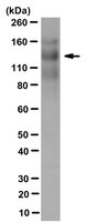
Detect Thrombospondin-1 using this Anti-Thrombospondin-1 Antibody, clone 133 validated for use in Western Blotting, Immunohistochemistry (Paraffin), Neutralizing, ELISA.
Key Applications
  • Western Blotting
  • Immunohistochemistry (Paraffin)
  • Neutralizing
  • ELISA
Application NotesWestern Blotting Analysis: A representative lot detected an upregulated thrombospondin-1 (TSP-1) expression in cultured primary rat vascular smooth muscle cells (VSMCs) upon treatment with either stainless steel (SS) ions or active TGF-β (Pallero, M.A., et al. (2010). J. Vasc. Res. 47(4):309-322).
Western Blotting Analysis: A representativev lot ditected full-length human thrombospondin-1 (hTSP-1) as well as hTSP-1 C-terminal fragments E3CaG (a.a. 648-1170), E3Ca (a.a. 648-945) and Ca (a.a. 692-945), but not hTSP-2, mouse TSP-1 (mTSP-1), or mTSP-2 by Western blotting under either reducing or non-reducing condition (Annis, D.S., et al. (2006). J. Thromb. Haemost. 4(2):459-468).
Western Blotting Analysis: A representative lot detected an upregulated thrombospondin-1 (TSP-1) in human pancreatic cancer Panc-1 cells upon siRNA-mediatted k-Ras knockdown (Fleming, J.B., et al. (2005). Mol. Cancer Res. 3(7):413-423).
Immunohistochemistry Analysis: A representative lot detected elevated thrombospondin-1 (TSP-1) immunoreactivity in vascular tissues surrounding stent materials due to in-stent restenosis (ISR) in paraffin-embedded sections of human coronary arteries from patients received either Taxus or Cypher drug-eluting stent implant (Pallero, M.A., et al. (2010). J. Vasc. Res. 47(4):309-322).
Neutralizing Analysis: Representative lots prevented alkaline-stripped human platelet thrombospondin-1 (sTSP-1) from activatating the latent form of TGF-beta secreted by cultured NMuMG murine mammary gland epithelial cells or bovine aortic endothelial (BAE) cells without affecting cellular signaling induced by the mature/active form of TGF-beta (Alcaraz, L.B., et al. (2014). J. Cell Biol. 205(3):409-428; Annis, D.S., et al. (2006). J. Thromb. Haemost. 4(2):459-468; Schultz-Cherry, S., and Murphy-Ullrich J.E. (1993). J. Cell Biol. 122(4):923-932).
Neutralizing Analysis: A representative lot inhibited stainless steel (SS) ions-induced TGF-β signaling in cultured primary rat vascular smooth muscle cells (VSMCs) as indicated by a suppressed induction of the synthetic/myofibroblastic protein ED-A FN (Pallero, M.A., et al. (2010). J. Vasc. Res. 47(4):309-322).
ELISA Analysis: A representativev lot ditected full-length human thrombospondin-1 (hTSP-1) as well as hTSP-1 C-terminal fragments E3CaG (a.a. 648-1170) and E3Ca (a.a. 648-945), but not hTSP-2, mouse TSP-1 (mTSP-1), or mTSP-2 by direct/non-sandwich ELISA (Annis, D.S., et al. (2006). J. Thromb. Haemost. 4(2):459-468).
Biological Information
ImmunogenAlkaline-stripped human platelet thrombospondin-1 (sTSP-1).
EpitopeCalcium-binding type-3 repeats.
Clone133
ConcentrationPlease refer to lot specific datasheet.
HostMouse
SpecificityClone 133 reacted with human thrombospondin-1 (hTSP-1), but not hTSP-2, mouse TSP-1 (mTSP-1), or mTSP-2. Clone 133 detected the C-terminal 50 kDa chymotryptic fragment of stripped TSP as well as recominant fragements containing the Calcium-binding type-3 repeats (a.a. 692-945) region (Annis, D.S., et al. (2006). J. Thromb. Haemost. 4(2):459-468; Schultz-Cherry, S., and Murphy-Ullrich J.E. (1993). J. Cell Biol. 122(4):923-932).
IsotypeIgG2bκ
Species Reactivity
  • Human
  • Rat
Antibody TypeMonoclonal Antibody
Entrez Gene Number
Gene Symbol
  • THBS
  • THBS1
  • TSP
  • TSP1
Purification MethodProtein G Purified
UniProt Number
Molecular Weight~129 kDa observed.
Physicochemical Information
Dimensions
Materials Information
Toxicological Information
Safety Information according to GHS
Safety Information
Product Usage Statements
Quality AssuranceEvaluated by Western Blotting in human thrombospondin-1 purified protein.

Western Blotting Analysis: 0.1 µg/mL of this antibody detected Thrombospondin-1 in 0.1 µg of human thrombospondin-1 purified protein.
Usage Statement
  • Unless otherwise stated in our catalog or other company documentation accompanying the product(s), our products are intended for research use only and are not to be used for any other purpose, which includes but is not limited to, unauthorized commercial uses, in vitro diagnostic uses, ex vivo or in vivo therapeutic uses or any type of consumption or application to humans or animals.
Storage and Shipping Information
Storage ConditionsStable for 1 year at -20°C from date of receipt.
Handling Recommendations: Upon receipt and prior to removing the cap, centrifuge the vial and gently mix the solution. Aliquot into microcentrifuge tubes and store at -20°C. Avoid repeated freeze/thaw cycles, which may damage IgG and affect product performance.
Packaging Information
Material Size100 μg
Transport Information
Supplemental Information
Specifications
Global Trade Item Number
Número de referencia GTIN
MABT879 04055977312973

Documentation

Anti-Thrombospondin-1 Antibody, clone 133 Ficha datos de seguridad (MSDS)

Título

Ficha técnica de seguridad del material (MSDS) 

Anti-Thrombospondin-1 Antibody, clone 133 Certificados de análisis

CargoNúmero de lote
Anti-Thrombospondin-1, clone 133 - 3197655 3197655
Anti-Thrombospondin-1, clone 133 - 3245598 3245598
Anti-Thrombospondin-1, clone 133 - 3429033 3429033
Anti-Thrombospondin-1, clone 133 - 4005668 4005668
Anti-Thrombospondin-1, clone 133 - 4084424 4084424
Anti-Thrombospondin-1, clone 133 - 4187028 4187028
Anti-Thrombospondin-1, clone 133 -Q2640174 Q2640174
Anti-Thrombospondin-1, clone 133 Monoclonal Antibody 2930954