Our broad portfolio consists of multiplex panels that allow you to choose, within the panel, analytes that best meet your needs. On a separate tab you can choose the premixed cytokine format or a single plex kit.
Cell Signaling Kits & MAPmates™
Choose fixed kits that allow you to explore entire pathways or processes. Or design your own kits by choosing single plex MAPmates™, following the provided guidelines.
The following MAPmates™ should not be plexed together:
-MAPmates™ that require a different assay buffer
-Phospho-specific and total MAPmate™ pairs, e.g. total GSK3β and GSK3β (Ser 9)
-PanTyr and site-specific MAPmates™, e.g. Phospho-EGF Receptor and phospho-STAT1 (Tyr701)
-More than 1 phospho-MAPmate™ for a single target (Akt, STAT3)
-GAPDH and β-Tubulin cannot be plexed with kits or MAPmates™ containing panTyr
.
Catalogue Number
Ordering Description
Qty/Pack
List
This item has been added to favorites.
Select A Species, Panel Type, Kit or Sample Type
To begin designing your MILLIPLEX® MAP kit select a species, a panel type or kit of interest.
Custom Premix Selecting "Custom Premix" option means that all of the beads you have chosen will be premixed in manufacturing before the kit is sent to you.
Catalogue Number
Ordering Description
Qty/Pack
List
This item has been added to favorites.
Species
Panel Type
Selected Kit
Qty
Catalogue Number
Ordering Description
Qty/Pack
List Price
96-Well Plate
Qty
Catalogue Number
Ordering Description
Qty/Pack
List Price
Add Additional Reagents (Buffer and Detection Kit is required for use with MAPmates)
Qty
Catalogue Number
Ordering Description
Qty/Pack
List Price
48-602MAG
Buffer Detection Kit for Magnetic Beads
1 Kit
Space Saver Option Customers purchasing multiple kits may choose to save storage space by eliminating the kit packaging and receiving their multiplex assay components in plastic bags for more compact storage.
This item has been added to favorites.
The Product Has Been Added To Your Cart
You can now customize another kit, choose a premixed kit, check out or close the ordering tool.
Attention: We have moved. Merck Millipore products are no longer available for purchase on MerckMillipore.com.Learn More
OP10L
Sigma-AldrichAnti-c-Myc (Ab-1) Mouse mAb (9E10)
This Anti-c-Myc (Ab-1) Mouse mAb (9E10) is validated for use in FC, Frozen Sections, Immunoblotting, IF, IP, Chromatin IP, Paraffin Sections for the detection of c-Myc (Ab-1).
More>>This Anti-c-Myc (Ab-1) Mouse mAb (9E10) is validated for use in FC, Frozen Sections, Immunoblotting, IF, IP, Chromatin IP, Paraffin Sections for the detection of c-Myc (Ab-1). Less<<
Anti-c-Myc (Ab-1) Mouse mAb (9E10) MSDS (material safety data sheet) or SDS, CoA and CoQ, dossiers, brochures and other available documents.
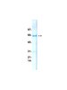
Recognizes a ~60-67 kDa c-Myc protein in HL60 cells and lung carcinoma tissue.
Catalogue Number
OP10L
Brand Family
Calbiochem®
References
References
LeGouy, E., et al. 1987. In Nuclear Oncogenes, Cold Spring Harbor Laboratory, 144. Cole, M.D. 1986. Ann. Rev. Gen.20, 361. Nisen, P.D., et al. 1986. Cancer Res.46, 6217. Nau, M.M., et al. 1985. Nature318, 69. Persson, H., et al. 1984. Science225, 687. Alitalo, K., et al. 1983. Proc. Natl. Acad. Sci. USA80, 1707.
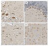
Flow Cytometry (20 µg/ml or use Cat. No. OP10F) Frozen Sections (2-10 µg/ml) Immunoblotting (1-5 µg/ml, see application references) Immunofluorescence (1-5 µg/ml or use Cat. No. OP10F) Immunoprecipitation (1 µg/sample) Chromatin Immunoprecipitation (5 µg/ml, see application references) Paraffin Sections (not recommended)
Application Comments
The c-Myc protein is extremely labile. c-Myc degradation can be as a result of freeze/thaw cycles, thus, we recommend using fresh lysates with a cocktail of protease inhibitors. c-Myc is often destroyed during the processing of paraffin sections and can therefore be difficult to detect in this application. This antibody recognizes the c-Myc protein and its cleavage products. The sequence of c-Myc predicts a ~45 kDa protein, but c-Myc migrates under reducing conditions as a ~64-67 kDa band. This antibody will not detect v-Myc but may react weakly to rodent c-Myc when used at high concentrations (10 µg/ml). An extra ~34-40 kDa band may be detected in a immunoblot. May also be used in immunofluorescence as well as the detection and purification of recombinant proteins tagged with the Myc epitope sequence EQKLISEEDL (see application references). The immunogen, c-Myc (Peptide-1) is also available for competition studies (Cat. No. PP06). Antibody should be titrated for optimal results in individual systems.
Biological Information
Immunogen
a synthetic peptide (AEEQKLISEEDLLRKRREQLKHKLEQLRNSCA) corresponding to amino acids 408-439 of human c-Myc
Immunogen
Human
Epitope
within amino acids 410-419
Clone
9E10
Host
Mouse
Isotype
IgG₁
Species Reactivity
Human
Mouse
Rat
Antibody Type
Monoclonal Antibody
Storage and Shipping Information
Ship Code
Ambient Temperature Only
Toxicity
Standard Handling
Storage
+2°C to +8°C
Do not freeze
Ok to freeze
Special Instructions
Reconstitute the lyophilized antibody with sterile PBS, pH 7.4, or sterile 20 mM Tris-saline (20 mM Tris containing 0.15 M NaCl), pH 7.4, to yield a final concentration of 100 µg/ml. Lyophilized antibodies should be resuspended at 4°C with occasional gentle mixing for at least 2 h.
Anti-c-Myc (Ab-1) Mouse mAb (9E10) Certificates of Analysis
Title
Lot Number
OP10L
References
Reference overview
LeGouy, E., et al. 1987. In Nuclear Oncogenes, Cold Spring Harbor Laboratory, 144. Cole, M.D. 1986. Ann. Rev. Gen.20, 361. Nisen, P.D., et al. 1986. Cancer Res.46, 6217. Nau, M.M., et al. 1985. Nature318, 69. Persson, H., et al. 1984. Science225, 687. Alitalo, K., et al. 1983. Proc. Natl. Acad. Sci. USA80, 1707.
Note that this data sheet is not lot-specific and is representative of the current specifications for this product. Please consult the vial label and the certificate of analysis for information on specific lots. Also note that shipping conditions may differ from storage conditions.
Revision
02-October-2007 RFH
Application
Flow Cytometry (20 µg/ml or use Cat. No. OP10F) Frozen Sections (2-10 µg/ml) Immunoblotting (1-5 µg/ml, see application references) Immunofluorescence (1-5 µg/ml or use Cat. No. OP10F) Immunoprecipitation (1 µg/sample) Chromatin Immunoprecipitation (5 µg/ml, see application references) Paraffin Sections (not recommended)
Description
Purified mouse monoclonal antibody generated by immunizing BALB/c mice with the specified immunogen and fusing splenocytes with SP2/0 mouse myeloma cells. Recognizes the ~64-67 kDa (apparent MW) c-Myc protein.
Background
The v-myc oncogene, initially identified in the MC29 avian retrovirus, causes myelocytomas, carcinomas, sarcomas and lymphomas, and belongs to a family of oncogenes conserved throughout evolution. In humans the family consists of five genes: c-myc, N-myc, R-myc, L-myc, and B-myc. Amplification of the c-myc gene has been found in several types of human tumors including lung, breast and colon carcinomas, while the N-myc gene has been found amplified in neuroblastomas, small cell lung carcinomas and Wilms tumors. L-myc is amplified in small cell lung cancer. Immunological studies have shown that the human c-myc gene gives rise to at least two nuclear phosphoproteins of ~64 kDa and ~67 kDa that exhibit relatively short (30 min) half lives in vivo and exhibit DNA binding properties in vitro.
Host
Mouse
Immunogen species
Human
Immunogen
a synthetic peptide (AEEQKLISEEDLLRKRREQLKHKLEQLRNSCA) corresponding to amino acids 408-439 of human c-Myc
Epitope
within amino acids 410-419
Clone
9E10
Isotype
IgG₁
Species
human, mouse (weakly), rat (weakly)
Positive control
HL-60 cells or lung carcinoma
Negative control
HT1080 cells
Form
Lyophilized
Formulation
Lyophilized from a volatile buffer, 100 µg BSA.
Preservative
None
Comments
The c-Myc protein is extremely labile. c-Myc degradation can be as a result of freeze/thaw cycles, thus, we recommend using fresh lysates with a cocktail of protease inhibitors. c-Myc is often destroyed during the processing of paraffin sections and can therefore be difficult to detect in this application. This antibody recognizes the c-Myc protein and its cleavage products. The sequence of c-Myc predicts a ~45 kDa protein, but c-Myc migrates under reducing conditions as a ~64-67 kDa band. This antibody will not detect v-Myc but may react weakly to rodent c-Myc when used at high concentrations (10 µg/ml). An extra ~34-40 kDa band may be detected in a immunoblot. May also be used in immunofluorescence as well as the detection and purification of recombinant proteins tagged with the Myc epitope sequence EQKLISEEDL (see application references). The immunogen, c-Myc (Peptide-1) is also available for competition studies (Cat. No. PP06). Antibody should be titrated for optimal results in individual systems.
Storage
+2°C to +8°C
Do Not Freeze
Ok to freeze
Special Instructions
Reconstitute the lyophilized antibody with sterile PBS, pH 7.4, or sterile 20 mM Tris-saline (20 mM Tris containing 0.15 M NaCl), pH 7.4, to yield a final concentration of 100 µg/ml. Lyophilized antibodies should be resuspended at 4°C with occasional gentle mixing for at least 2 h.
Toxicity
Standard Handling
References
LeGouy, E., et al. 1987. In Nuclear Oncogenes, Cold Spring Harbor Laboratory, 144. Cole, M.D. 1986. Ann. Rev. Gen.20, 361. Nisen, P.D., et al. 1986. Cancer Res.46, 6217. Nau, M.M., et al. 1985. Nature318, 69. Persson, H., et al. 1984. Science225, 687. Alitalo, K., et al. 1983. Proc. Natl. Acad. Sci. USA80, 1707.
Application references
Epitope Tagging
Munro, S., et al. 1986. Cell46, 291.
Immunoblotting
Evan, G.I., et al. 1985. Mol. Cell. Biol.5, 3610.
Chromatin Immunoprecipitation
Sanyal, K., et al. 2004. Proc. Natl. Acad. Sci. USA101, 11374.