An effective freezing/thawing method for human pluripotent stem cells cultured in chemically-defined and feeder-free conditions.
Nishishita, N; Muramatsu, M; Kawamata, S
American journal of stem cells
4
38-49
2015
Show Abstract
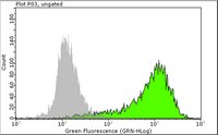
Culturing human Pluripotent Stem Cells (hPSC)s in chemically defined medium and feeder-free condition can facilitate metabolome and proteome analysis of culturing cells and medium, and reduce regulatory concerns for clinical application of cells. And in addition, if hPSC are passaged and cryopreserved in single cells it also facilitates quality control of cells at single cell level. Here we report a robust single cell freezing and thawing method of hPSCs cultured in chemically-defined medium TeSR(TM)-E8(TM) and on cost-effective recombinant human Vitronectin-N (rhVTN-N)-coated dish. Cells are dissociated into single cells with recombinant TrypLE(TM) Select and 0.5 mM EDTA/PBS (3:1 solution) in the presence of Rock inhibitor and cryopreserved with chemically defined CryoStem(TM). Approximately 60% of cells were viable after dissociation. Aggrewell(TM) 400 was used to form cell clumps of 500 cells after thaw in the presence of Rock inhibitor and cells were cultured for two days with TeSR-E8. Cells clumps were then seeded on rhVTN-N-coated dish and cultured with TeSR-E8 for two days prior to the first passage after thawing. Number of viable cells at the first passage increased around 10 times of that just before freezing. This robust single cell freezing method for hPSCs cultured in chemically defined medium will facilitate quality control of cultured cells at single cell level before cryopreservation and consequently assure the quality of cells in frozen vials for further manipulation after thawing. | | | 25973330
 |
V-myc immortalizes human neural stem cells in the absence of pluripotency-associated traits.
Pino-Barrio, MJ; García-García, E; Menéndez, P; Martínez-Serrano, A
PloS one
10
e0118499
2015
Show Abstract
A better understanding of the molecular mechanisms governing stem cell self-renewal will foster the use of different types of stem cells in disease modeling and cell therapy strategies. Immortalization, understood as the capacity for indefinite expansion, is needed for the generation of any cell line. In the case of v-myc immortalized multipotent human Neural Stem Cells (hNSCs), we hypothesized that v-myc immortalization could induce a more de-differentiated state in v-myc hNSC lines. To test this, we investigated the expression of surface, biochemical and genetic markers of stemness and pluripotency in v-myc immortalized and control hNSCs (primary precursors, that is, neurospheres) and compared these two cell types to human Embryonic Stem Cells (hESCs) and fibroblasts. Using a Hierarchical Clustering method and a Principal Component Analysis (PCA), the v-myc hNSCs associated with their counterparts hNSCs (in the absence of v-myc) and displayed a differential expression pattern when compared to hESCs. Moreover, the expression analysis of pluripotency markers suggested no evidence supporting a reprogramming-like process despite the increment in telomerase expression. In conclusion, v-myc expression in hNSC lines ensures self-renewal through the activation of some genes involved in the maintenance of stem cell properties in multipotent cells but does not alter the expression of key pluripotency-associated genes. | | | 25764185
|
Derivation of induced pluripotent stem cells from orangutan skin fibroblasts.
Ramaswamy, K; Yik, WY; Wang, XM; Oliphant, EN; Lu, W; Shibata, D; Ryder, OA; Hacia, JG
BMC research notes
8
577
2015
Show Abstract
Orangutans are an endangered species whose natural habitats are restricted to the Southeast Asian islands of Borneo and Sumatra. Along with the African great apes, orangutans are among the closest living relatives to humans. For potential species conservation and functional genomics studies, we derived induced pluripotent stem cells (iPSCs) from cryopreserved somatic cells obtained from captive orangutans.Primary skin fibroblasts from two Sumatran orangutans were transduced with retroviral vectors expressing the human OCT4, SOX2, KLF4, and c-MYC factors. Candidate orangutan iPSCs were characterized by global gene expression and DNA copy number analysis. All were consistent with pluripotency and provided no evidence of large genomic insertions or deletions. In addition, orangutan iPSCs were capable of producing cells derived from all three germ layers in vitro through embryoid body differentiation assays and in vivo through teratoma formation in immune-compromised mice.We demonstrate that orangutan skin fibroblasts are capable of being reprogrammed into iPSCs with hallmark molecular signatures and differentiation potential. We suggest that reprogramming orangutan somatic cells in genome resource banks could provide new opportunities for advancing assisted reproductive technologies relevant for species conservation efforts. Furthermore, orangutan iPSCs could have applications for investigating the phenotypic relevance of genomic changes that occurred in the human, African great ape, and/or orangutan lineages. This provides opportunities for orangutan cell culture models that would otherwise be impossible to develop from living donors due to the invasive nature of the procedures required for obtaining primary cells. | | | 26475477
|
Alveolar epithelial differentiation of human induced pluripotent stem cells in a rotating bioreactor.
Ghaedi, M; Mendez, JJ; Bove, PF; Sivarapatna, A; Raredon, MS; Niklason, LE
Biomaterials
35
699-710
2014
Show Abstract
Traditional stem cell differentiation protocols make use of a variety of cytokines including growth factors (GFs) and inhibitors in an effort to provide appropriate signals for tissue specific differentiation. In this study, iPSC-derived type II pneumocytes (iPSC-ATII) as well as native isolated human type II pneumocytes (hATII) were differentiated toward a type I phenotype using a unique air-liquid interface (ALI) system that relies on a rotating apparatus that mimics in vivo respiratory conditions. A relatively homogenous population of alveolar type II-like cells from iPSC was first generated (iPSC-ATII cells), which had phenotypic properties similar to mature human alveolar type II cells. iPSC-ATII cells were then cultured in a specially designed rotating culture apparatus. The effectiveness of the ALI bioreactor was compared with the effectiveness of small molecule-based differentiation of type II pneumocytes toward type 1 pneumocytes. The dynamics of differentiation were examined by quantitative reverse transcriptase polymerase chain reaction (qRT-PCR), flow cytometry and immunocytochemistry. iPSC-ATII and hATII cells cultured in the ALI bioreactor had higher levels of type I markers, including aquaporin-5(AQ5), caveolin-1, and T1α, at both the RNA and protein levels as compared with the flask-grown iPSC-ATII and hATII that had been treated with small molecules to induce differentiation. In summary, this study demonstrates that a rotating bioreactor culture system that provides an air-liquid interface is a potent inducer of type I epithelial differentiation for both iPS-ATII cells and hATII cells, and provides a method for large-scale production of alveolar epithelium for tissue engineering and drug discovery. | Immunocytochemistry | | 24144903
|
Patient-specific naturally gene-reverted induced pluripotent stem cells in recessive dystrophic epidermolysis bullosa.
Tolar, J; McGrath, JA; Xia, L; Riddle, MJ; Lees, CJ; Eide, C; Keene, DR; Liu, L; Osborn, MJ; Lund, TC; Blazar, BR; Wagner, JE
The Journal of investigative dermatology
134
1246-54
2014
Show Abstract
Spontaneous reversion of disease-causing mutations has been observed in some genetic disorders. In our clinical observations of severe generalized recessive dystrophic epidermolysis bullosa (RDEB), a currently incurable blistering genodermatosis caused by loss-of-function mutations in COL7A1 that results in a deficit of type VII collagen (C7), we have observed patches of healthy-appearing skin on some individuals. When biopsied, this skin revealed somatic mosaicism resulting in the self-correction of C7 deficiency. We believe this source of cells could represent an opportunity for translational 'natural' gene therapy. We show that revertant RDEB keratinocytes expressing functional C7 can be reprogrammed into induced pluripotent stem cells (iPSCs) and that self-corrected RDEB iPSCs can be induced to differentiate into either epidermal or hematopoietic cell populations. Our results give proof-of-principle that an inexhaustible supply of functional patient-specific revertant cells can be obtained--potentially relevant to local wound therapy and systemic hematopoietic cell transplantation. This technology may also avoid some of the major limitations of other cell therapy strategies, e.g., immune rejection and insertional mutagenesis, which are associated with viral- and nonviral-mediated gene therapy. We believe this approach should be the starting point for autologous cellular therapies using 'natural' gene therapy in RDEB and other diseases. | | | 24317394
|
Human oocytes reprogram adult somatic nuclei of a type 1 diabetic to diploid pluripotent stem cells.
Yamada, Mitsutoshi, et al.
Nature, (2014)
2014
Show Abstract
The transfer of somatic cell nuclei into oocytes can give rise to pluripotent stem cells that are consistently equivalent to embryonic stem cells, holding promise for autologous cell replacement therapy. Although methods to induce pluripotent stem cells from somatic cells by transcription factors are widely used in basic research, numerous differences between induced pluripotent stem cells and embryonic stem cells have been reported, potentially affecting their clinical use. Because of the therapeutic potential of diploid embryonic stem-cell lines derived from adult cells of diseased human subjects, we have systematically investigated the parameters affecting efficiency of blastocyst development and stem-cell derivation. Here we show that improvements to the oocyte activation protocol, including the use of both kinase and translation inhibitors, and cell culture in the presence of histone deacetylase inhibitors, promote development to the blastocyst stage. Developmental efficiency varied between oocyte donors, and was inversely related to the number of days of hormonal stimulation required for oocyte maturation, whereas the daily dose of gonadotropin or the total number of metaphase II oocytes retrieved did not affect developmental outcome. Because the use of concentrated Sendai virus for cell fusion induced an increase in intracellular calcium concentration, causing premature oocyte activation, we used diluted Sendai virus in calcium-free medium. Using this modified nuclear transfer protocol, we derived diploid pluripotent stem-cell lines from somatic cells of a newborn and, for the first time, an adult, a female with type 1 diabetes. | | | 24776804
|
Derivation of transgene-free human induced pluripotent stem cells from human peripheral T cells in defined culture conditions.
Kishino, Y; Seki, T; Fujita, J; Yuasa, S; Tohyama, S; Kunitomi, A; Tabei, R; Nakajima, K; Okada, M; Hirano, A; Kanazawa, H; Fukuda, K
PloS one
9
e97397
2014
Show Abstract
Recently, induced pluripotent stem cells (iPSCs) were established as promising cell sources for revolutionary regenerative therapies. The initial culture system used for iPSC generation needed fetal calf serum in the culture medium and mouse embryonic fibroblast as a feeder layer, both of which could possibly transfer unknown exogenous antigens and pathogens into the iPSC population. Therefore, the development of culture systems designed to minimize such potential risks has become increasingly vital for future applications of iPSCs for clinical use. On another front, although donor cell types for generating iPSCs are wide-ranging, T cells have attracted attention as unique cell sources for iPSCs generation because T cell-derived iPSCs (TiPSCs) have a unique monoclonal T cell receptor genomic rearrangement that enables their differentiation into antigen-specific T cells, which can be applied to novel immunotherapies. In the present study, we generated transgene-free human TiPSCs using a combination of activated human T cells and Sendai virus under defined culture conditions. These TiPSCs expressed pluripotent markers by quantitative PCR and immunostaining, had a normal karyotype, and were capable of differentiating into cells from all three germ layers. This method of TiPSCs generation is more suitable for the therapeutic application of iPSC technology because it lowers the risks associated with the presence of undefined, animal-derived feeder cells and serum. Therefore this work will lead to establishment of safer iPSCs and extended clinical application. | | | 24824994
|
Cell-autonomous correction of ring chromosomes in human induced pluripotent stem cells.
Bershteyn, M; Hayashi, Y; Desachy, G; Hsiao, EC; Sami, S; Tsang, KM; Weiss, LA; Kriegstein, AR; Yamanaka, S; Wynshaw-Boris, A
Nature
507
99-103
2014
Show Abstract
Ring chromosomes are structural aberrations commonly associated with birth defects, mental disabilities and growth retardation. Rings form after fusion of the long and short arms of a chromosome, and are sometimes associated with large terminal deletions. Owing to the severity of these large aberrations that can affect multiple contiguous genes, no possible therapeutic strategies for ring chromosome disorders have been proposed. During cell division, ring chromosomes can exhibit unstable behaviour leading to continuous production of aneuploid progeny with low viability and high cellular death rate. The overall consequences of this chromosomal instability have been largely unexplored in experimental model systems. Here we generated human induced pluripotent stem cells (iPSCs) from patient fibroblasts containing ring chromosomes with large deletions and found that reprogrammed cells lost the abnormal chromosome and duplicated the wild-type homologue through the compensatory uniparental disomy (UPD) mechanism. The karyotypically normal iPSCs with isodisomy for the corrected chromosome outgrew co-existing aneuploid populations, enabling rapid and efficient isolation of patient-derived iPSCs devoid of the original chromosomal aberration. Our results suggest a fundamentally different function for cellular reprogramming as a means of 'chromosome therapy' to reverse combined loss-of-function across many genes in cells with large-scale aberrations involving ring structures. In addition, our work provides an experimentally tractable human cellular system for studying mechanisms of chromosomal number control, which is of critical relevance to human development and disease. | Immunofluorescence | | 24413397
|
Development of a high-yield technique to isolate spermatogonial stem cells from porcine testes.
Park, MH; Park, JE; Kim, MS; Lee, KY; Park, HJ; Yun, JI; Choi, JH; Lee, Es; Lee, ST
Journal of assisted reproduction and genetics
31
983-91
2014
Show Abstract
To date, the methods available for isolating spermatogonial stem cells (SSCs) from porcine testicular cells have a low efficiency of cell separating. Therefore, we tried to develop a novel isolation technique with a high-yield cell separating ability to isolate SSCs from porcine testes.We confirmed the presence of SSCs by measuring alkaline phosphatase (AP) activity and SSC-specific gene expression in neonatal porcine testis-derived testicular cells. Subsequently, the isolation of SSCs from testicular cells was performed using different techniques as follows: differential plating (DP), double DP, Petri dish plating post-DP, magnetic-activated cell sorting (MACS), and MACS post-DP. Positive AP staining was used to assess and compare the isolation efficiency of each method.Petri dish plating post-DP resulted in the highest isolation efficiency. The putative SSCs isolated using this method was then further characterized by analyzing the expression of SSC-specific genes and -related proteins, and germ cell-specific genes. OCT4, NANOG, EPCAM, THY1, and UCHL1 were expressed transcriptionally, and OCT4, NANOG, SOX2, TRA-1-60, TRA-1-81, and PLZF were expressed translationally in 86 % of the isolated SSCs. In contrast, no difference was observed in the percentage of cells expressing luteinizing hormone receptor (LHR), a Leydig cell-specific protein, or GATA4, a Sertoli cell-specific protein, between SSCs and negative control cells. In addition, transcriptional expression of VASA, a primordial germ cell-specific marker, and DAZL, a premeiotic germ cell-specific marker, wasn't and was detected, respectively.We successfully developed a novel high-yield technique to isolate SSCs from porcine testes to facilitate future porcine SSC-related research. | Fluorescence Activated Cell Sorting (FACS), Immunocytochemistry | | 24938360
|
Generation and characterization of leukemia inhibitory factor-dependent equine induced pluripotent stem cells from adult dermal fibroblasts.
Whitworth, DJ; Ovchinnikov, DA; Sun, J; Fortuna, PR; Wolvetang, EJ
Stem cells and development
23
1515-23
2014
Show Abstract
In this study we have reprogrammed dermal fibroblasts from an adult female horse into equine induced pluripotent stem cells (equiPSCs). These equiPSCs are dependent only on leukemia inhibitory factor (LIF), placing them in striking contrast to previously derived equiPSCs that have been shown to be co-dependent on both LIF and basic fibroblast growth factor (bFGF). These equiPSCs have a normal karyotype and have been maintained beyond 60 passages. They possess alkaline phosphatase activity and express eqNANOG, eqOCT4, and eqTERT mRNA. Immunocytochemistry confirmed that they produce NANOG, REX1, SSEA4, TRA1-60, and TRA1-81. While our equiPSCs are LIF dependent, bFGF co-stimulates their proliferation via the PI3K/AKT pathway. EquiPSCs lack expression of eqXIST and immunostaining for H3K27me3, suggesting that during reprogramming the inactive X chromosome has likely been reactivated to generate cells that have two active X chromosomes. EquiPSCs form embryoid bodies and in vitro teratomas that contain derivatives of all three germ layers. These LIF-dependent equiPSCs likely reflect a more naive state of pluripotency than equiPSCs that are co-dependent on both LIF and bFGF and so provide a novel resource for understanding pluripotency in the horse. | | | 24555755
|