Loss of the AE3 Cl(-)/HCO(-) 3 exchanger in mice affects rate-dependent inotropy and stress-related AKT signaling in heart.
Prasad, V; Lorenz, JN; Lasko, VM; Nieman, ML; Al Moamen, NJ; Shull, GE
Frontiers in physiology
4
399
2013
Show Abstract
Cl(-)/HCO(-) 3 exchangers are expressed abundantly in cardiac muscle, suggesting that HCO(-) 3 extrusion serves an important function in heart. Mice lacking Anion Exchanger Isoform 3 (AE3), a major cardiac Cl(-)/HCO(-) 3 exchanger, appear healthy, but loss of AE3 causes decompensation in a hypertrophic cardiomyopathy (HCM) model. Using intra-ventricular pressure analysis, in vivo pacing, and molecular studies we identified physiological and biochemical changes caused by loss of AE3 that may contribute to decompensation in HCM. AE3-null mice had normal cardiac contractility under basal conditions and after β-adrenergic stimulation, but pacing of hearts revealed that frequency-dependent inotropy was blunted, suggesting that AE3-mediated HCO(-) 3 extrusion is required for a robust force-frequency response (FFR) during acute biomechanical stress in vivo. Modest changes in expression of proteins that affect Ca(2+)-handling were observed, but Ca(2+)-transient analysis of AE3-null myocytes showed normal twitch-amplitude and Ca(2+)-clearance. Phosphorylation and expression of several proteins implicated in HCM and FFR, including phospholamban (PLN), myosin binding protein C, and troponin I were not altered in hearts of paced AE3-null mice; however, phosphorylation of Akt, which plays a central role in mechanosensory signaling, was significantly higher in paced AE3-null hearts than in wild-type controls and phosphorylation of AMPK, which is affected by Akt and is involved in energy metabolism and some cases of HCM, was reduced. These data show loss of AE3 leads to impaired rate-dependent inotropy, appears to affect mechanical stress-responsive signaling, and reduces activation of AMPK, which may contribute to decompensation in heart failure. | | | 24427143
 |
The 10-20-30 training concept improves performance and health profile in moderately trained runners.
Gunnarsson, TP; Bangsbo, J
Journal of applied physiology (Bethesda, Md. : 1985)
113
16-24
2012
Show Abstract
The effect of an alteration from regular endurance to interval (10-20-30) training on the health profile, muscular adaptations, maximum oxygen uptake (Vo(2max)), and performance of runners was examined. Eighteen moderately trained individuals (6 females and 12 males; Vo(2max): 52.2 ± 1.5 ml·kg(-1)·min(-1)) (means ± SE) were divided into a high-intensity training (10-20-30; 3 women and 7 men) and a control (CON; 3 women and 5 men) group. For a 7-wk intervention period the 10-20-30 replaced all training sessions with 10-20-30 training consisting of low-, moderate-, and high-speed running (less than 30%, less than 60%, and greater than 90% of maximal intensity) for 30, 20, and 10 s, respectively, in three or four 5-min intervals interspersed by 2 min of recovery, reducing training volume by 54% (14.0 ± 0.9 vs. 30.4 ± 2.3 km/wk) while CON continued the normal training. After the intervention period Vo(2max) in 10-20-30 was 4% higher, and performance in a 1,500-m and a 5-km run improved (P less than 0.05) by 21 and 48 s, respectively. In 10-20-30, systolic blood pressure was reduced (P less than 0.05) by 5 ± 2 mmHg, and total and low-density lipoprotein (LDL) cholesterol was lowered (P less than 0.05) by 0.5 ± 0.2 and 0.4 ± 0.1 mmol/l, respectively. No alterations were observed in CON. Muscle membrane proteins and enzyme activity did not change in either of the groups. The present study shows that interval training with short 10-s near-maximal bouts can improve performance and Vo(2max) despite a ∼50% reduction in training volume. In addition, the 10-20-30 training regime lowers resting systolic blood pressure and blood cholesterol, suggesting a beneficial effect on the health profile of already trained individuals. | | | 22556401
|
Four weeks of normobaric "live high-train low" do not alter muscular or systemic capacity for maintaining pH and K⁺ homeostasis during intense exercise.
Nordsborg, NB; Siebenmann, C; Jacobs, RA; Rasmussen, P; Diaz, V; Robach, P; Lundby, C
Journal of applied physiology (Bethesda, Md. : 1985)
112
2027-36
2012
Show Abstract
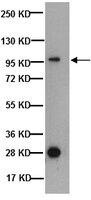
It was investigated if athletes subjected to 4 wk of living in normobaric hypoxia (3,000 m; 16 h/day) while training at 800-1,300 m ["live high-train low" (LHTL)] increase muscular and systemic capacity for maintaining pH and K(+) homeostasis as well as intense exercise performance. The design was double-blind and placebo controlled. Mean power during 30-s all-out cycling was similar before and immediately after LHTL (650 ± 31 vs. 628 ± 32 W; n = 10) and placebo exposure (658 ± 22 vs. 660 ± 23 W; n = 6). Supporting the performance data, arterial plasma pH, lactate, and K(+) during submaximal and maximal exercise were also unaffected by the intervention in both groups. In addition, muscle buffer capacity (in mmol H(+)·kg dry wt(-1)·pH(-1)) was similar before and after in both the LHTL (140 ± 12 vs. 140 ± 16) and placebo group (145 ± 2 vs. 140 ± 3). The expression of sarcolemmal H(+) transporters (Na(+)/H(+) exchanger 1, monocarboxylate transporters 1 and 4), as well as expression of Na(+)-K(+) pump subunits-α(1), -α(2), and -β(1) was also similar before and after the intervention. In conclusion, muscular and systemic capacity for maintaining pH and K(+) balance during exercise is similar before and after 4 wk of placebo-controlled normobaric LHTL. In accordance, 30-s all-out sprint ability was similar before and after LHTL. | Western Blotting | Human | 22461443
|
Protein Phosphatase 2A (B55{alpha}) Prevents Premature Activation of Forkhead Transcription Factor FoxM1 by Antagonizing Cyclin A/ Cyclin-dependent Kinase
Alvarez-Fernández M, Halim VA, Aprelia M, Mohammed S, Medema RH
The Journal of biological chemistry
286
33029-36. Epub 2011 Aug 3.
2011
Show Abstract
The forkhead transcription factor FoxM1 controls expression of a large number of genes that are specifically expressed during the G(2) phase of the cell cycle. Throughout most of the cell cycle, FoxM1 activity is restrained by an autoinhibitory mechanism, involving a repressor domain present in the N-terminal part of the protein. Activation of FoxM1 in G(2) is achieved by Cyclin A/Cyclin-dependent kinase (Cdk)-mediated phosphorylation, which alleviates autoinhibition by the N-terminal repressor domain. Here, we show that FoxM1 interacts with B55α, a regulatory subunit of protein phosphatase 2A (PP2A). B55α binds the catalytic subunit of PP2A, and this promotes dephosphorylation and inactivation of FoxM1. Indeed, we find that overexpression of B55α results in decreased FoxM1 activity. Inversely, depletion of B55α results in premature activation of FoxM1. The activation of FoxM1 that is observed upon depletion of B55α is fully dependent on Cyclin A/Cdk-mediated phosphorylation of FoxM1. Taken together, these data demonstrate that B55α acts to antagonize Cyclin A/Cdk-dependent activation of FoxM1, to ensure that FoxM1 activity is restricted to the G(2) phase of the cell cycle. | | | 21813648
|
Cortactin phosphorylation regulates cell invasion through a pH-dependent pathway.
Magalhaes, MA; Larson, DR; Mader, CC; Bravo-Cordero, JJ; Gil-Henn, H; Oser, M; Chen, X; Koleske, AJ; Condeelis, J
The Journal of cell biology
195
903-20
2011
Show Abstract
Invadopodia are invasive protrusions with proteolytic activity uniquely found in tumor cells. Cortactin phosphorylation is a key step during invadopodia maturation, regulating Nck1 binding and cofilin activity. The precise mechanism of cortactin-dependent cofilin regulation and the roles of this pathway in invadopodia maturation and cell invasion are not fully understood. We provide evidence that cortactin-cofilin binding is regulated by local pH changes at invadopodia that are mediated by the sodium-hydrogen exchanger NHE1. Furthermore, cortactin tyrosine phosphorylation mediates the recruitment of NHE1 to the invadopodium compartment, where it locally increases the pH to cause the release of cofilin from cortactin. We show that this mechanism involving cortactin phosphorylation, local pH increase, and cofilin activation regulates the dynamic cycles of invadopodium protrusion and retraction and is essential for cell invasion in 3D. Together, these findings identify a novel pH-dependent regulation of cell invasion. | | | 22105349
|
Silencing of cardiac mitochondrial NHE1 prevents mitochondrial permeability transition pore opening.
Villa-Abrille, MC; Cingolani, E; Cingolani, HE; Alvarez, BV
American journal of physiology. Heart and circulatory physiology
300
H1237-51
2011
Show Abstract
Inhibition of Na(+)/H(+) exchanger 1 (NHE1) reduces cardiac ischemia-reperfusion (I/R) injury and also cardiac hypertrophy and failure. Although the mechanisms underlying these NHE1-mediated effects suggest delay of mitochondrial permeability transition pore (MPTP) opening, and reduction of mitochondrial-derived superoxide production, the possibility of NHE1 blockade targeting mitochondria has been incompletely explored. A short-hairpin RNA sequence mediating specific knock down of NHE1 expression was incorporated into a lentiviral vector (shRNA-NHE1) and transduced in the rat myocardium. NHE1 expression of mitochondrial lysates revealed that shRNA-NHE1 transductions reduced mitochondrial NHE1 (mNHE1) by ∼60%, supporting the expression of NHE1 in mitochondria membranes. Electron microscopy studies corroborate the presence of NHE1 in heart mitochondria. Immunostaining of rat cardiomyocytes also suggests colocalization of NHE1 with the mitochondrial marker cytochrome c oxidase. To examine the functional role of mNHE1, mitochondrial suspensions were exposed to increasing concentrations of CaCl(2) to induce MPTP opening and consequently mitochondrial swelling. shRNA-NHE1 transduction reduced CaCl(2)-induced mitochondrial swelling by 64 ± 4%. Whereas the NHE1 inhibitor HOE-642 (10 μM) decreased mitochondrial Ca(2+)-induced swelling in rats transduced with nonsilencing RNAi (37 ± 6%), no additional HOE-642 effects were detected in mitochondria from rats transduced with shRNA-NHE1. We have characterized the expression and function of NHE1 in rat heart mitochondria. Because mitochondria from rats injected with shRNA-NHE1 present a high threshold for MPTP formation, the beneficial effects of NHE1 inhibition in I/R resulting from mitochondrial targeting should be considered. | | | 21297023
|
The spatial organization of proton and lactate transport in a rat brain tumor.
Grillon, E; Farion, R; Fablet, K; De Waard, M; Tse, CM; Donowitz, M; Rémy, C; Coles, JA
PloS one
6
e17416
2011
Show Abstract
Tumors create a heterogeneous acidic microenvironment which assists their growth and which must be taken into account in the design of drugs and their delivery. In addition, the acidic extracellular pH (pHe) is itself exploited in several experimental techniques for drug delivery. The way the acidity is created is not clear. We report here the spatial organization of key proton-handling proteins in C6 gliomas in rat brain. The mean profiles across the tumor rim of the Na+/H+ exchanger NHE1, and the lactate-H+ cotransporter MCT1, both showed peaks. NHE1, which is important for extension and migration of cells in vitro, showed a peak 1.55 times higher than in extratumoural tissue at 0.33 mm from the edge. MCT1 had a broader peak, further into the tumor (maximum 1.76 fold at 1.0 mm from the edge). In contrast, MCT4 and the carbonic anhydrase CAIX, which are associated with hypoxia, were not significantly upregulated in the rim. The spatial distribution of MCT4 was highly correlated with that of CAIX, suggesting that their expression is regulated by the same factors. Since protons extruded by NHE1 diffuse away through extracellular clefts, NHE1 requires a continuous source of intracellular protons. From the stoichiometries of metabolic pathways that produce or consume H+, and the greater availability of glucose compared to oxygen in most parts of a tumor, we support the classic view that most of the net proton efflux from C6 gliomas originates in glycolytic formation of lactate and H+ inside the tumor, but add that some lactate is taken up into cells in the rim on MCT1, and some lactate diffuses away, leaving its associated protons available to re-enter cells for extrusion on NHE1. Therapeutic inhibition of NHE1, MCT1 or CAIX is predicted to affect different parts of a tumor. | | | 21390324
|
Relationship between performance at different exercise intensities and skeletal muscle characteristics.
Iaia, FM; Perez-Gomez, J; Thomassen, M; Nordsborg, NB; Hellsten, Y; Bangsbo, J
Journal of applied physiology (Bethesda, Md. : 1985)
110
1555-63
2011
Show Abstract
The hypothesis investigated whether exercise performance over a broad range of intensities is determined by specific skeletal muscle characteristics. Seven subjects performed 8-10 exhaustive cycle trials at different workloads, ranging from 150 to 700 W (150 min to 20 s). No relationships between the performance times at high and low workloads were observed. A relationship (P less than 0.05) was noticed between the percentage of fast-twitch x fibers and the exercise time at 579 ± 21 W (∼30 s; r(2) = 0.88). Capillary-to-fiber-ratio (r(2): 0.58-0.85) was related (P less than 0.05) to exercise time at work intensities ranging from 395 to 270 W (2.5-21 min). Capillary density was correlated (r(2) = 0.68; P less than 0.05) with the net rate of plasma K(+) accumulation during an ∼3-min bout and was estimated to explain 50-80% (P less than 0.05) of the total variance observed in exercise performances lasting ∼30 s to 3 min. The Na(+)-K(+) pump β(1)-subunit expression was found to account for 13-34% (P less than 0.05) during exhaustive exercise of ∼1-4 min. In conclusion, exercise performance at different intensities is related to specific physiological variables. A large distribution of fast-twitch x fibers may play a role during very intense efforts, i.e., ∼30 s. Muscle capillaries and the Na(+)-K(+) pump β(1)-subunit seem to be important determinants for performance during exhaustive high-intensity exercises lasting between 30 s and 4 min. | | | 21436467
|
Exercise-induced regulation of muscular Na+-K+ pump, FXYD1, and NHE1 mRNA and protein expression: importance of training status, intensity, and muscle type.
Rasmussen, MK; Juel, C; Nordsborg, NB
American journal of physiology. Regulatory, integrative and comparative physiology
300
R1209-20
2011
Show Abstract
It is investigated if exercise-induced mRNA changes cause similar protein expression changes of Na(+)-K(+) pump isoforms (α(1), α(2), β(1), β(2)), FXYD1, and Na(+)/K(+) exchanger (NHE1) in rat skeletal muscle. Expression was evaluated (n = 8 per group) in soleus and extensor digutorum longus after 1 day, 3 days, and 3 wk (5 sessions/wk) of either sprint (4 × 3-min sprint + 1-min rest) or endurance (20 min) running. Two hours after exercise on day 1, no change in protein expression was apparent in either training group or muscle, whereas sprint exercise increased the mRNA of soleus α(2) (4.9 ± 0.8-fold; P less than 0.05), β(2) (13.2 ± 4.4-fold; P less than 0.001), and NHE1 (12.0 ± 3.1-fold; P less than 0.01). Two hours after sprint exercise, protein expression normalized to control samples was higher on day 3 than day 1 for soleus α(1) (41 ± 18% increase vs. 15 ± 8% reduction; P less than 0.05), α(2) (64 ± 35% increase vs. 37 ± 12% reduction; P less than 0.05), β(1) (17 ± 21% increase vs. 14 ± 29% reduction; P less than 0.05), and FXYD1 (35 ± 16% increase vs. 13 ± 10% reduction; P less than 0.05). In contrast, on day 3, soleus α(1) (0.1 ± 0.1-fold; P less than 0.001), α(2) (0.2 ± 0.1-fold; P less than 0.001), β(1) (0.4 ± 0.1-fold; P less than 0.05), and β(2)-mRNA (2.9 ± 1.7-fold; P less than 0.001) expression was lower than after exercise on day 1. After 3 wk of training, no change in protein expression relative to control existed. In conclusion, increased expression of Na(+)-K(+) pump subunits, FXYD1 and NHE1 after 3 days exercise training does not appear to be an effect of increased constitutive mRNA levels. Importantly, sprint exercise can reduce mRNA expression concomitant with increased protein expression. | | | 21325644
|
Angiotensin II-mediated biphasic regulation of proximal tubular Na+/H+ exchanger 3 is impaired during oxidative stress.
Banday, AA; Lokhandwala, MF
American journal of physiology. Renal physiology
301
F364-70
2011
Show Abstract
Angiotensin (ANG) II via AT1 receptors (AT1Rs) maintains sodium homeostasis by regulating renal sodium transporters including Na(+)/H(+) exchanger 3 (NHE3) in a biphasic manner. Low-ANG II concentration stimulates whereas high concentrations inhibit NHE3 activity. Oxidative stress has been shown to upregulate AT1R function that could modulate the ANG II-mediated NHE3 regulation. This study was designed to identify the signaling pathways responsible for ANG II-mediated biphasic regulation of proximal tubular NHE3 and the effect of oxidative stress on this phenomenon. Male Sprague-Dawley rats were chronically treated with a pro-oxidant L-buthionine sulfoximine (BSO) with and without an antioxidant tempol in tap water for 3 wk. BSO-treated rats exhibited oxidative stress and high blood pressure. At low concentration (1 pM) ANG II increased NHE3 activity in proximal tubules from all animals. However, in BSO-treated rats, the stimulation was more robust and was normalized by tempol treatment. ANG II (1 pM)-mediated NHE3 activation was abolished by AT1R blocker, intracellular Ca(2+) chelator, and inhibitors of phospholipase C (PLC) and Ca(2+)-dependent calmodulin (CaM) but it was insensitive to Giα and protein kinase C inhibitors or AT2R antagonist. A high concentration of ANG II (1 μM) inhibited NHE3 activity in control and tempol-treated rats. However, in BSO-treated rats, ANG II (1 μM) continued to induce NHE3 stimulation. Tempol restored the inhibitory effect of ANG II (1 μM) in BSO-treated rats. The inhibitory effect of ANG II (1 μM) involved AT1R-dependent, cGMP-dependent protein kinase (PKG) activation and was independent of AT2 receptor and nitric oxide signaling. We conclude that ANG II stimulates NHE3 via AT1R-PLC-CaM pathway and inhibits NHE3 by AT1R-PKG activation. Oxidative stress impaired ANG II-mediated NHE3 biphasic response in that stimulation was observed at both high- and low-ANG II concentration. | Western Blotting | | 21593187
|