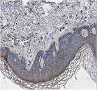
Decorin is processed by three isoforms of bone morphogenetic protein-1 (BMP1).
von Marschall, Z; Fisher, LW
Biochemical and biophysical research communications
391
1374-8
2010
Show Abstract
The secreted small proteoglycan, decorin, modulates collagen fibril formation as well as the bioactivity of various members of the transforming growth factor-beta (TGFbeta) superfamily. Indeed, recombinant prodecorin has been used in several gene therapy experiments to inhibit unwanted fibrosis in model diseases of the kidney, heart, and other tissues although the status of the propeptide within the target tissues is unknown. Currently the protease that removes the highly conserved propeptide from decorin is unproven. Using a variety of approaches, we show that three isoforms of the Tolloid-related bone morphogenetic protein-1 (BMP1) can effectively remove the propeptide from human prodecorin resulting in the well-established mature proteoglycan. Classic BMP1, the full-length gene transcript mTLD (BMP1-3), and BMP1-5 (isoform lacking the CUB3 domain thought to be important for efficient type I collagen C-propeptidase activity) all removed the analogous propeptides from both recombinant human prodecorin and murine probiglycan. Furthermore, the timed removal of the propeptide was found to not be necessary for the addition of decorin's single glycosaminoglycan chain. Decorin therefore joins the growing list of matrix and bioactive molecules processed/activated by the BMP1/Tolloid family. Since the third member of the Class I small leucine-rich proteooglycan (SLRP) superfamily, asporin, also contains a similar cleavage motif at the appropriate location, we propose that the removal of these propeptides by members of the BMP1 family is an additional characteristic of Class I SLRP. | 20026052
 |
Expression and localization of the two small proteoglycans biglycan and decorin in developing human skeletal and non-skeletal tissues.
Bianco, P; Fisher, LW; Young, MF; Termine, JD; Robey, PG
The journal of histochemistry and cytochemistry : official journal of the Histochemistry Society
38
1549-63
1990
Show Abstract
The messenger RNAs and core proteins of the two small chondroitin/dermatan sulfate proteoglycans, biglycan and decorin, were localized in developing human bone and other tissues by both 35S-labeled RNA probes and antibodies directed against synthetic peptides corresponding to nonhomologous regions of the two core proteins. Biglycan and decorin expression and localization were substantially divergent and sometimes mutually exclusive. In developing bones, spatially restricted patterns of gene expression and/or matrix localization of the two proteoglycans were identified in articular regions, epiphyseal cartilage, vascular canals, subperichondral regions, and periosteum, and indicated the association of each molecule with specific developmental events at specific sites. Study of non-skeletal tissues revealed that decorin was associated with all major type I (and type II) collagen-rich connective tissues. Conversely, biglycan was expressed and localized in a range of specialized cell types, including connective tissue (skeletal myofibers, endothelial cells) and epithelial cells (differentiating keratinocytes, renal tubular epithelia). Biglycan core protein was localized at the cell surface of certain cell types (e.g., keratinocytes). Whereas the distribution of decorin was consistent with matrix-centered functions, possibly related to regulation of growth of collagen fibers, the distribution of biglycan pointed to other function(s), perhaps related to cell regulation. | 2212616
 |


