Millipore Sigma Vibrant Logo

MABF2163-25UG Anti-CD33 Antibody, clone P67.6

View Products on Sigmaaldrich.com
MABF2163-25UG
25 μg  
Retrieving price...
Price could not be retrieved
Minimum Quantity is a multiple of
Maximum Quantity is
Upon Order Completion More Information
You Saved ()
 
Request Pricing
Fulfillment and delivery delayed
In Stock 
Discontinued
Limited Quantities Available
Availability to be confirmed
    Remaining : Will advise
      Remaining : Will advise
      Will advise
      Contact Customer Service
      Contact Customer Service


      Contact Customer Service

      Overview

      Replacement Information

      Key Spec Table

      Species ReactivityKey ApplicationsHostFormatAntibody Type
      HFC, ICC, InhibitionMPurifiedMonoclonal Antibody
      Description
      Catalogue NumberMABF2163-25UG
      DescriptionAnti-CD33 Antibody, clone P67.6
      Alternate Names
      • Myeloid cell surface antigen CD33
      • Sialic acid-binding Ig-like lectin 3
      • Siglec-3
      • gp67
      Background InformationMyeloid cell surface antigen CD33 (UniProt: P20138; also known as Sialic acid-binding Ig-like lectin 3, Siglec-3, gp67, CD33) is encoded by the CD33 (also known as SIGLEC3) gene (Gene ID: 945) in human. CD33 is a single-pass type I membrane sialoadhesion protein that is expressed by myeloid stem cells, myeloblasts, monoblasts, monocytes, macrophages, and granulocyte precursors and preferentially binds to alpha-2,6-linked sialic acid. CD33 is not expressed in erythrocytes, platelets, B-cells, T-cells, and NK cells. Hence, it can serve as a marker for myeloid cells. It is synthesized with a signal peptide (aa 1-17), which is subsequently cleaved off in the mature form. It contains an extracellular domain (aa 18-259), a transmembrane domain (aa 260-282), and a cytoplasmic domain (aa 283-364). CD33 contains two copies of a cytoplasmic motif (aa 338-343 and 356-361), which is referred to as the immunoreceptor tyrosine-based inhibitor motif (ITIM). This motif is involved in modulation of cellular responses. In the immune response, CD33 may act as an inhibitory receptor upon ligand induced tyrosine phosphorylation by recruiting cytoplasmic phosphatase(s) via their SH2 domain(s) that block signal transduction through dephosphorylation of signaling molecules. Human CD33 is reported to contain two tyrosine residues in its cytoplasmic domain (tyrosine 340 and 358) that when phosphorylated could function as docking sites for the phosphatases, SHP-1 and/or SHP-2, and enable CD33 to function as an inhibitory receptor.
      References
      Product Information
      FormatPurified
      PresentationPurified mouse monoclonal antibody IgG1 in PBS without azide.
      Applications
      ApplicationAnti-CD33, clone P67.6, Cat. No. MABF2163, is a mouse monoclonal antibody that detects CD33 and has been tested for use in Flow Cytometry, Immunocytochemistry, and Inhibition assays.
      Key Applications
      • Flow Cytometry
      • Immunocytochemistry
      • Inhibition
      Application NotesInhibition Assays: A representative lot of CD33 conjugated to calicheamicin inhibited inhibits the formation of colonies from AML marrow samples. (Hamann, P.R., et. al. (2002). Bioconjug Chem. 13(1):47-58).

      Flow Cytometry Analysis: A representative lot detected CD33 in Flow Cytometry applications (Kussick, S.J., et. al. (2003). Arch Pathol Lab Med. 127(9):1140-7).

      Immunocytochemistry Analysis: A representative lot detected CD33 in Immunocytochemistry applications (van Der Velden, V.H., et. al. (2001). Blood. 97(10):3197-204).
      Biological Information
      ImmunogenFMY9S5 cells expressing CD33.
      CloneP67.6
      ConcentrationPlease refer to lot specific datasheet.
      HostMouse
      SpecificityClone P67.6 specifically detects human CD 33.
      IsotypeIgG1κ
      Species Reactivity
      • Human
      Species Reactivity NoteHuman.
      Antibody TypeMonoclonal Antibody
      Entrez Gene Number
      Gene Symbol
      • CD33
      • SIGLEC3
      Purification MethodProtein G purified
      UniProt Number
      Molecular Weight39.82 kDa calculated.
      Physicochemical Information
      Dimensions
      Materials Information
      Toxicological Information
      Safety Information according to GHS
      Safety Information
      Product Usage Statements
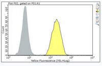
      Quality AssuranceEvaluated by Flow Cytometry in THP-1 cells.

      Flow Cytometry Analysis: 1 µg of this antibody detected CD33 in one milliion THP-1 cells.
      Usage Statement
      • Unless otherwise stated in our catalog or other company documentation accompanying the product(s), our products are intended for research use only and are not to be used for any other purpose, which includes but is not limited to, unauthorized commercial uses, in vitro diagnostic uses, ex vivo or in vivo therapeutic uses or any type of consumption or application to humans or animals.
      Storage and Shipping Information
      Storage ConditionsStable for 1 year at -20°C from date of receipt. Handling Recommendations: Upon receipt and prior to removing the cap, centrifuge the vial and gently mix the solution. Aliquot into microcentrifuge tubes and store at -20°C. Avoid repeated freeze/thaw cycles, which may damage IgG and affect product performance.
      Packaging Information
      Material Size25 μg
      Transport Information
      Supplemental Information
      Specifications
      Global Trade Item Number
      Catalogue Number GTIN
      MABF2163-25UG 04054839551147