Wenn Sie das Fenster schließen, wird Ihre Konfiguration nicht gespeichert, es sei denn, Sie haben Ihren Artikel in die Bestellung aufgenommen oder zu Ihren Favoriten hinzugefügt.
Klicken Sie auf OK, um das MILLIPLEX® MAP-Tool zu schließen oder auf Abbrechen, um zu Ihrer Auswahl zurückzukehren.
Wählen Sie konfigurierbare Panels & Premixed-Kits - ODER - Kits für die zelluläre Signaltransduktion & MAPmates™
Konfigurieren Sie Ihre MILLIPLEX® MAP-Kits und lassen sich den Preis anzeigen.
Konfigurierbare Panels & Premixed-Kits
Unser breites Angebot enthält Multiplex-Panels, für die Sie die Analyten auswählen können, die am besten für Ihre Anwendung geeignet sind. Unter einem separaten Register können Sie das Premixed-Cytokin-Format oder ein Singleplex-Kit wählen.
Kits für die zelluläre Signaltransduktion & MAPmates™
Wählen Sie gebrauchsfertige Kits zur Erforschung gesamter Signalwege oder Prozesse. Oder konfigurieren Sie Ihre eigenen Kits mit Singleplex MAPmates™.
Die folgenden MAPmates™ sollten nicht zusammen analysiert werden: -MAPmates™, die einen unterschiedlichen Assaypuffer erfordern. -Phosphospezifische und MAPmate™ Gesamtkombinationen wie Gesamt-GSK3β und Gesamt-GSK3β (Ser 9). -PanTyr und locusspezifische MAPmates™, z.B. Phospho-EGF-Rezeptor und Phospho-STAT1 (Tyr701). -Mehr als 1 Phospho-MAPmate™ für ein einziges Target (Akt, STAT3). -GAPDH und β-Tubulin können nicht mit Kits oder MAPmates™, die panTyr enthalten, analysiert werden.
.
Bestellnummer
Bestellinformationen
St./Pkg.
Liste
Dieser Artikel wurde zu Ihren Favoriten hinzugefügt.
Wählen Sie bitte Spezies, Panelart, Kit oder Probenart
Um Ihr MILLIPLEX® MAP-Kit zu konfigurieren, wählen Sie zunächst eine Spezies, eine Panelart und/oder ein Kit.
Custom Premix Selecting "Custom Premix" option means that all of the beads you have chosen will be premixed in manufacturing before the kit is sent to you.
Catalogue Number
Ordering Description
Qty/Pack
List
Dieser Artikel wurde zu Ihren Favoriten hinzugefügt.
Spezies
Panelart
Gewähltes Kit
Menge
Bestellnummer
Bestellinformationen
St./Pkg.
Listenpreis
96-Well Plate
Menge
Bestellnummer
Bestellinformationen
St./Pkg.
Listenpreis
Weitere Reagenzien hinzufügen (MAPmates erfordern die Verwendung eines Puffer- und Detektionskits)
Menge
Bestellnummer
Bestellinformationen
St./Pkg.
Listenpreis
48-602MAG
Buffer Detection Kit for Magnetic Beads
1 Kit
Platzsparende Option Kunden, die mehrere Kits kaufen, können ihre Multiplex-Assaykomponenten in Kunststoffbeuteln anstelle von Packungen erhalten, um eine kompaktere Lagerung zu ermöglichen.
Dieser Artikel wurde zu Ihren Favoriten hinzugefügt.
Das Produkt wurde in Ihre Bestellung aufgenommen
Sie können nun ein weiteres Kit konfigurieren, ein Premixed-Kit wählen, zur Kasse gehen oder das Bestell-Tool schließen.
SCC245
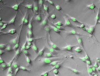
Sigma-AldrichYUMMER1.7-H2B-GFP5 Mouse Melanoma Cell Line
The YUMMER1.7-H2B-GFP5 mouse melanoma cell line is a valuable model for studies of immune checkpoint inhibition and mechanisms of anti-tumor responses.
More>>The YUMMER1.7-H2B-GFP5 mouse melanoma cell line is a valuable model for studies of immune checkpoint inhibition and mechanisms of anti-tumor responses. Less<<
Empfohlene Produkte
Übersicht
Replacement Information
Description
Catalogue Number
SCC245
Description
YUMMER1.7-H2B-GFP5 Mouse Melanoma Cell Line
Overview
The YUMMER1.7 H2B-GFP mouse melanoma cell line is both immunocompetent and reflective of the somatic mutations common in melanomas. The YUMMER1.7 H2B-GFP cell line carries three driver mutations of melanoma: Braf V600E, Pten -/- and Cdkn2 -/- (1,2). In addition, the YUMMER1.7 H2B-GFP cell line harbors a high frequency of stable UV-induced somatic mutations which have been shown to stimulate host adaptive immune response (3). YUMMER1.7-H2B-GFP cells stably express GFP fused to histone H2B, allowing visualization of chromosomal dynamics and facilitating in vivo detection of YUMMER1.7 melanoma cells and YUMMER1.7-derived tumors. The unique features of the YUMMER1.7 H2B-GFP cell line make it a valuable model for studies of immune checkpoint inhibition and mechanisms of anti-tumor responses.
Source: The YUMMER1.7 H2B-GFP mouse melanoma cell line is a tetraploid clonal isolate derived from YUMM1.7 cells exposed to UVB radiation into which a histone H2B-GFP lentiviral fusion construct has been stably introduced via transduction (4). The original YUMM1.7 cell line was derived from a 4-hydroxytamoxifen-induced melanoma tumor in a male C57/B1/6 mouse into which mutations from the Braf/Pten genetically-engineered mouse model had been introduced via backcrossing (1). The YUMMER1.7 H2B-GFP cell line harbors the Braf V600E mutation and is homozygous negative for wild-type Pten and Cdkn2 (3).
Alternate Names
Yale University Mouse Melanoma Exposed to Radiation 1.7
Background Information
The great promise of immune-based therapies in cancer and recent progress in successful application of these approaches has brought to the fore the necessity of immune-competent models to evaluate immune system responses to cancer cells. Melanomas exhibit relatively high somatic mutation burden, and these mutations can act as neoantigens that generate anti-tumor immune responses. The development of immunocompetent cell models is critical to the advancement of cancer immunotherapy and understanding of immune responses, although few tractable model systems are available.
References: 1. Meeth K et al. (2016) The YUMM lines: a series of congenic mouse melanoma cell lines with defined genetic alterations. Pigment Cell Melanoma Res 29(5): 590-597. 2. Dankort D et al. (2009) Braf(V600E) cooperates with Pten loss to induce metastatic melanoma. Nat Genet. 41(5): 544-552. 3. Wang J et al. (2017) UV-induced somatic mutations elicit a functional T cell response in the YUMMER1.7 mouse melanoma model. Pigment Cell Melanoma Res 30(4): 428-435. 4. Zhou T et al. (2020) IL-18BP is a secreted immune checkpoint and barrier to IL-18 immunotherapy Nature 583(7817):609-614.
References
Product Information
Applications
Application
The YUMMER1.7-H2B-GFP5 mouse melanoma cell line is a valuable model for studies of immune checkpoint inhibition and mechanisms of anti-tumor responses.
Key Applications
Cell Culture
Application Notes
This product is intended for sale and sold solely to academic institutions for internal academic research use per the terms of the “Academic Use Agreement” as detailed in the product documentation. For information regarding any other use, please contact licensing@emdmillipore.com.
Biological Information
Cell Line Type
Cancer Cells
Physicochemical Information
Dimensions
Materials Information
Toxicological Information
Safety Information according to GHS
Safety Information
Product Usage Statements
Quality Assurance
• Each vial contains ≥ 1X10⁶ viable cells. • Cells are tested negative for infectious diseases by a Mouse Essential CLEAR panel by Charles River Animal Diagnostic Services. • Cells are verified to be of mouse origin and negative for inter-species contamination from rat, chinese hamster, Golden Syrian hamster, human and non-human primate (NHP) as assessed by a Contamination CLEAR panel by Charles River Animal Diagnostic Services. • Cells are negative for mycoplasma contamination.
Usage Statement
This product contains genetically modified organisms (GMO). Within the EU GMOs are regulated by Directives 2001/18/EC and 2009/41/EC of the European Parliament and of the Council and their national implementation in the member States respectively. This legislation obliges Merck to request certain information about you and the establishment where the GMOs are being handled. Click here for Enduser Declaration (EUD) Form.
Unless otherwise stated in our catalog or other company documentation accompanying the product(s), our products are intended for research use only and are not to be used for any other purpose, which includes but is not limited to, unauthorized commercial uses, in vitro diagnostic uses, ex vivo or in vivo therapeutic uses or any type of consumption or application to humans or animals.
Storage and Shipping Information
Storage Conditions
Store in liquid nitrogen. The cells can be cultured for at least 10 passages after initial thawing without significantly affecting the cell marker expression and functionality.