Wenn Sie das Fenster schließen, wird Ihre Konfiguration nicht gespeichert, es sei denn, Sie haben Ihren Artikel in die Bestellung aufgenommen oder zu Ihren Favoriten hinzugefügt.
Klicken Sie auf OK, um das MILLIPLEX® MAP-Tool zu schließen oder auf Abbrechen, um zu Ihrer Auswahl zurückzukehren.
Wählen Sie konfigurierbare Panels & Premixed-Kits - ODER - Kits für die zelluläre Signaltransduktion & MAPmates™
Konfigurieren Sie Ihre MILLIPLEX® MAP-Kits und lassen sich den Preis anzeigen.
Konfigurierbare Panels & Premixed-Kits
Unser breites Angebot enthält Multiplex-Panels, für die Sie die Analyten auswählen können, die am besten für Ihre Anwendung geeignet sind. Unter einem separaten Register können Sie das Premixed-Cytokin-Format oder ein Singleplex-Kit wählen.
Kits für die zelluläre Signaltransduktion & MAPmates™
Wählen Sie gebrauchsfertige Kits zur Erforschung gesamter Signalwege oder Prozesse. Oder konfigurieren Sie Ihre eigenen Kits mit Singleplex MAPmates™.
Die folgenden MAPmates™ sollten nicht zusammen analysiert werden: -MAPmates™, die einen unterschiedlichen Assaypuffer erfordern. -Phosphospezifische und MAPmate™ Gesamtkombinationen wie Gesamt-GSK3β und Gesamt-GSK3β (Ser 9). -PanTyr und locusspezifische MAPmates™, z.B. Phospho-EGF-Rezeptor und Phospho-STAT1 (Tyr701). -Mehr als 1 Phospho-MAPmate™ für ein einziges Target (Akt, STAT3). -GAPDH und β-Tubulin können nicht mit Kits oder MAPmates™, die panTyr enthalten, analysiert werden.
.
Bestellnummer
Bestellinformationen
St./Pkg.
Liste
Dieser Artikel wurde zu Ihren Favoriten hinzugefügt.
Wählen Sie bitte Spezies, Panelart, Kit oder Probenart
Um Ihr MILLIPLEX® MAP-Kit zu konfigurieren, wählen Sie zunächst eine Spezies, eine Panelart und/oder ein Kit.
Custom Premix Selecting "Custom Premix" option means that all of the beads you have chosen will be premixed in manufacturing before the kit is sent to you.
Catalogue Number
Ordering Description
Qty/Pack
List
Dieser Artikel wurde zu Ihren Favoriten hinzugefügt.
Spezies
Panelart
Gewähltes Kit
Menge
Bestellnummer
Bestellinformationen
St./Pkg.
Listenpreis
96-Well Plate
Menge
Bestellnummer
Bestellinformationen
St./Pkg.
Listenpreis
Weitere Reagenzien hinzufügen (MAPmates erfordern die Verwendung eines Puffer- und Detektionskits)
Menge
Bestellnummer
Bestellinformationen
St./Pkg.
Listenpreis
48-602MAG
Buffer Detection Kit for Magnetic Beads
1 Kit
Platzsparende Option Kunden, die mehrere Kits kaufen, können ihre Multiplex-Assaykomponenten in Kunststoffbeuteln anstelle von Packungen erhalten, um eine kompaktere Lagerung zu ermöglichen.
Dieser Artikel wurde zu Ihren Favoriten hinzugefügt.
Das Produkt wurde in Ihre Bestellung aufgenommen
Sie können nun ein weiteres Kit konfigurieren, ein Premixed-Kit wählen, zur Kasse gehen oder das Bestell-Tool schließen.
SCC623
Sigma-AldrichMIN6 Mouse Insulinoma Cell Line
The MIN6 cell line is a long-term stable mouse pancreatic beta-cell line that retains characteristics of mature β-cells and can be used to analyze insulin secretion/sensitivity in diabetes research.
More>>The MIN6 cell line is a long-term stable mouse pancreatic beta-cell line that retains characteristics of mature β-cells and can be used to analyze insulin secretion/sensitivity in diabetes research. Less<<
Empfohlene Produkte
Übersicht
Replacement Information
Description
Catalogue Number
SCC623
Description
MIN6 Mouse Insulinoma Cell Line
Alternate Names
Min6
MIN-6
Mouse INsulinoma 6
Min6 cells
Background Information
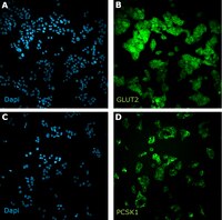
The Mouse Insulinoma Cell Line (MIN6), was derived from an insulinoma tissue that developed in a transgenic mouse in which the expression of the Simian Virus 40 Large T antigen was driven by the human insulin promoter.1,2,3 In comparison to the primary pancreatic b-cells, MIN6 thrives reliably and far more robustly in cell culture and yet retains the important characteristics of the b-cells, such as the exclusive expression of the so-called “liver-type” glucose transporter (Glut2) and their ability to modulate the insulin secretion in response to the changing glucose concentration in the growth medium, and it has been a staple strain in diabetes research since its establishment in 1990.1
Additionally, the expression of the major histocompatibility complex I (MHC I) is lacking in MIN6, as in the non-diabetic b-cells, but can be induced by pro-inflammatory cytokines,1,4,5 which would render them susceptible to T-cell mediated cytotoxicity. This closely mimics the behavior of the b-cells isolated from diabetic tissues, where chronic inflammation is commonly observed, and it has made MIN6 an attractive cell line model for investigating the mechanism of the b-cells loss due to their autoimmune destruction.
References 1. Miyazaki J, Araki K, Yamato E, Ikegami H, Asano T, Shibasaki Y, Oka Y, Yamamura K. 1990. Establishment of a pancreatic beta cell line that retains glucose-inducible insulin secretion: special reference to expression of glucose transporter isoforms. Endocrinology. 27(1):126-32. 2. Sarvetnick N, Liggitt D, Pitts SL, Hansen SE, Stewart TA. 1988. Insulin-dependent diabetes mellitus induced in transgenic mice by ectopic expression of class II MHC and interferon-gamma. Cell. 52(5):773-82. Pytynia KB, Dahlstrom KR, Sturgis EM. 2014. Epidemiology of HPV-associated oropharyngeal cancer. Oral Oncol. 50(5): 380-6. 3. Ullrich A, Dull TJ, Gray A, Philips JA 3rd, Peter S. 1982. Variation in the sequence and modification state of the human insulin gene flanking regions. Nucleic Acids Res. 10(7):2225-40. 4. Jiang H, Li Y, Shen M, Liang Y, Qian Y, Dai H, Xu K, Xu X, Lv H, Zhang J, Yang T, Fu Q. 2022. Interferon-α promotes MHC I antigen presentation of islet β cells through STAT1-IRF7 pathway in type 1 diabetes. Immunology. 166(2):210-221. 5. Javeed N, Her TK, Brown MR, Vanderboom P, Rakshit K, Egan AM, Vella A, Lanza I, Matveyenko AV. 2021. Pro-inflammatory β cell small extracellular vesicles induce β cell failure through activation of the CXCL10/CXCR3 axis in diabetes. Cell Rep. 36(8):109613.
The MIN6 cell line is a long-term stable mouse pancreatic beta-cell line that retains characteristics of mature β-cells and can be used to analyze insulin secretion/sensitivity in diabetes research.
Key Applications
Cell Culture
Application Notes
This product is intended for sale and sold solely to academic institutions for internal academic research use per the terms of the “Academic Use Agreement” as detailed in the product documentation. For information regarding any other use, please contact licensing@emdmillipore.com.
Biological Information
Cell Line Type
Cancer Cells
Pancreas
Physicochemical Information
Dimensions
Materials Information
Toxicological Information
Safety Information according to GHS
Safety Information
Product Usage Statements
Quality Assurance
• Cells are verified to be of mouse origin and negative for human, rat, Chinese hamster, Golden Syrian hamster, and non-human primate interspecies contamination, as assessed by a Contamination Clear panel by Charles River Animal Diagnostic Services • Cells tested negative for infectious diseases against a Mouse Essential CLEAR panel by Charles River Animal Diagnostic Services. • Cells tested negative for mycoplasma.
Usage Statement
Unless otherwise stated in our catalog or other company documentation accompanying the product(s), our products are intended for research use only and are not to be used for any other purpose, which includes but is not limited to, unauthorized commercial uses, in vitro diagnostic uses, ex vivo or in vivo therapeutic uses or any type of consumption or application to humans or animals.
This product contains genetically modified organisms (GMO). Within the EU GMOs are regulated by Directives 2001/18/EC and 2009/41/EC of the European Parliament and of the Council and their national implementation in the member States respectively. This legislation obliges Merck to request certain information about you and the establishment where the GMOs are being handled. Click here for Enduser Declaration (EUD) Form.
Storage and Shipping Information
Storage Conditions
The cells should be stored in liquid nitrogen. The cells can be cultured for at least 10 passages after initial thawing without significantly affecting the cell marker expression and functionality.