Wenn Sie das Fenster schließen, wird Ihre Konfiguration nicht gespeichert, es sei denn, Sie haben Ihren Artikel in die Bestellung aufgenommen oder zu Ihren Favoriten hinzugefügt.
Klicken Sie auf OK, um das MILLIPLEX® MAP-Tool zu schließen oder auf Abbrechen, um zu Ihrer Auswahl zurückzukehren.
Wählen Sie konfigurierbare Panels & Premixed-Kits - ODER - Kits für die zelluläre Signaltransduktion & MAPmates™
Konfigurieren Sie Ihre MILLIPLEX® MAP-Kits und lassen sich den Preis anzeigen.
Konfigurierbare Panels & Premixed-Kits
Unser breites Angebot enthält Multiplex-Panels, für die Sie die Analyten auswählen können, die am besten für Ihre Anwendung geeignet sind. Unter einem separaten Register können Sie das Premixed-Cytokin-Format oder ein Singleplex-Kit wählen.
Kits für die zelluläre Signaltransduktion & MAPmates™
Wählen Sie gebrauchsfertige Kits zur Erforschung gesamter Signalwege oder Prozesse. Oder konfigurieren Sie Ihre eigenen Kits mit Singleplex MAPmates™.
Die folgenden MAPmates™ sollten nicht zusammen analysiert werden: -MAPmates™, die einen unterschiedlichen Assaypuffer erfordern. -Phosphospezifische und MAPmate™ Gesamtkombinationen wie Gesamt-GSK3β und Gesamt-GSK3β (Ser 9). -PanTyr und locusspezifische MAPmates™, z.B. Phospho-EGF-Rezeptor und Phospho-STAT1 (Tyr701). -Mehr als 1 Phospho-MAPmate™ für ein einziges Target (Akt, STAT3). -GAPDH und β-Tubulin können nicht mit Kits oder MAPmates™, die panTyr enthalten, analysiert werden.
.
Bestellnummer
Bestellinformationen
St./Pkg.
Liste
Dieser Artikel wurde zu Ihren Favoriten hinzugefügt.
Wählen Sie bitte Spezies, Panelart, Kit oder Probenart
Um Ihr MILLIPLEX® MAP-Kit zu konfigurieren, wählen Sie zunächst eine Spezies, eine Panelart und/oder ein Kit.
Custom Premix Selecting "Custom Premix" option means that all of the beads you have chosen will be premixed in manufacturing before the kit is sent to you.
Catalogue Number
Ordering Description
Qty/Pack
List
Dieser Artikel wurde zu Ihren Favoriten hinzugefügt.
Spezies
Panelart
Gewähltes Kit
Menge
Bestellnummer
Bestellinformationen
St./Pkg.
Listenpreis
96-Well Plate
Menge
Bestellnummer
Bestellinformationen
St./Pkg.
Listenpreis
Weitere Reagenzien hinzufügen (MAPmates erfordern die Verwendung eines Puffer- und Detektionskits)
Menge
Bestellnummer
Bestellinformationen
St./Pkg.
Listenpreis
48-602MAG
Buffer Detection Kit for Magnetic Beads
1 Kit
Platzsparende Option Kunden, die mehrere Kits kaufen, können ihre Multiplex-Assaykomponenten in Kunststoffbeuteln anstelle von Packungen erhalten, um eine kompaktere Lagerung zu ermöglichen.
Dieser Artikel wurde zu Ihren Favoriten hinzugefügt.
Das Produkt wurde in Ihre Bestellung aufgenommen
Sie können nun ein weiteres Kit konfigurieren, ein Premixed-Kit wählen, zur Kasse gehen oder das Bestell-Tool schließen.
LentiBrite™ GFP-HMGB1 Lentiviral Biosensor
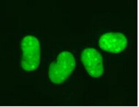
: SDB (Sicherheitsdatenblätter), Analysenzertifikate und Qualitätszertifikate, Dossiers, Broschüren und andere verfügbare Dokumente.
Read our application note in Nature Methods! http://www.nature.com/app_notes/nmeth/2012/121007/pdf/an8620.pdf (Click Here!)
Learn more about the advantages of our LentiBrite Lentiviral Biosensors! Click Here
Biosensors can be used to detect the presence/absence of a particular protein as well as the subcellular location of that protein within the live state of a cell. Fluorescent tags are often desired as a means to visualize the protein of interest within a cell by either fluorescent microscopy or time-lapse video capture. Visualizing live cells without disruption allows researchers to observe cellular conditions in real time.
Lentiviral vector systems are a popular research tool used to introduce gene products into cells. Lentiviral transfection has advantages over non-viral methods such as chemical-based transfection including higher-efficiency transfection of dividing and non-dividing cells, long-term stable expression of the transgene, and low immunogenicity.
EMD Millipore is introducing LentiBrite™ Lentiviral Biosensors, a new suite of pre-packaged lentiviral particles encoding important and foundational proteins of autophagy, apoptosis, and cell structure for visualization under different cell/disease states in live cell and in vitro analysis.
Pre-packaged, fluorescently-tagged with GFP & RFP
Higher efficiency transfection as compared to traditional chemical-based and other non-viral-based transfection methods
Ability to transfect dividing, non-dividing, and difficult-to-transfect cell types, such as primary cells or stem cells
Non-disruptive towards cellular function
EMD Millipore’s LentiBrite™ GFP-HMGB1 lentiviral particles provide bright fluorescence and precise localization to enable live cell analysis of HMGB1 translocation in difficult-to-transfect cell types.
Alternate Names
High mobility group protein B1
High mobility group protein 1
Background Information
HMBG1 (high mobility box group 1) functions in the nucleus of most cells to stabilize nucleosomes and enhance binding of transcription factor complexes to DNA. In certain activated inflammatory cells, HMGB1 is actively secreted, whereas necrotic cells passively release HMGB1. In both cases, extracellular HMGB1 can bind to multiple immune cell types via RAGE (receptor for advanced glycation end products) to stimulate chemotaxis and cytokine production, which, depending on the context, results in inflammation, wound healing, or immunogenic tumor cell death. Cells undergoing autophagy also display translocation of HMGB1, from the nucleus to the cytoplasm. Expression of genetic fusions between HMGB1 and fluorescent proteins permits monitoring of HMGB1 translocation in real time. EMD Millipore’s LentiBrite™ GFP-HMGB1 lentiviral particles provide bright fluorescence and precise localization to enable live cell analysis of HMGB1 translocation in difficult-to-transfect cell types.
References
Product Information
Components
TagGFP2-HMGB Lentivirus: One vial containing 25 µL of lentiviral particles at a minimum of 3 x 10E8 infectious units (IFU) per mL. For lot-specific titer information, please see lot specific “Viral Titer” in the product specifications of the datasheet.
Promoter EF-1 (Elongation Factor-1)
Multiplicty of Infection (MOI) MOI = Ratio of # of infectious lentiviral particles (IFU) to # of cells being infected. Typical MOI values for high transduction efficiency and signal intensity are in the range of 20-40. For this target, some cell types may require lower MOIs (e.g., HT-1080, HeLa, U2OS), while others may require higher MOIs (e.g., human umbilical vein endothelial cells (HUVEC), human mesenchymal stem cells (HuMSC)). NOTE: MOI should be titrated and optimized by the end user for each cell type and lentiviral target to achieve desired transduction efficiency and signal intensity.
Fluorescence Microscopy Imaging: (See Figure 1 in datasheet) U2OS cells were plated in a chamber slide and transduced with lentiviral particles at an MOI of 10 for 24 hours. After media replacement and 48 hours further incubation, cells were fixed with formaldehyde and mounted. Images were obtained by oil immersion wide-field fluorescence microscopy. The GFP-HMGB1 displays in the nucleus of cells.
Immunocytochemistry Comparison and Inhibitor Analysis: (See Figure 2 in datasheet) Similar to Figure 1 (See datasheet), HeLa cells were plated in a chamber slide and transduced with lentiviral particles at an MOI of 10 for 24 hours. After 24 hours, media was replaced and cells were then further incubated for 48 hours. Immunocytochemical staining (blue) of the same fields of view with a nuclear stain (DAPI) reveals similar expression patterns to the protein-GFP (green).
Difficult-to-transfect Cell Types: (See Figure 3 in datasheet) Primary cell types HUVEC and Human mesenchymal stem cells (HuMSC) were plated in chamber slides and transduced with lentiviral particles at an MOI of 20 for 24 hours.
For optimal fluorescent visualization, it is recommended to analyze the target expression level within 24-48 hrs after transfection/infection for optimal live cell analysis, as fluorescent intensity may dim over time, especially in difficult-to-transfect cell lines. Infected cells may be frozen down after successful transfection/infection and thawed in culture to retain positive fluorescent expression beyond 24-48 hrs. Length and intensity of fluorescent expression varies between cell lines. Higher MOIs may be required for difficult-to-transfect cell lines.
Evaluated by transduction of HT-1080 cells and fluorescent imaging performed for assessment of target localization and transduction efficiency.
Usage Statement
This product contains genetically modified organisms (GMO). Within the EU GMOs are regulated by Directives 2001/18/EC and 2009/41/EC of the European Parliament and of the Council and their national implementation in the member States respectively. This legislation obliges Merck to request certain information about you and the establishment where the GMOs are being handled. Click here for Enduser Declaration (EUD) Form.
Unless otherwise stated in our catalog or other company documentation accompanying the product(s), our products are intended for research use only and are not to be used for any other purpose, which includes but is not limited to, unauthorized commercial uses, in vitro diagnostic uses, ex vivo or in vivo therapeutic uses or any type of consumption or application to humans or animals.
Storage and Shipping Information
Storage Conditions
Storage and Handling Lentivirus is stable for at least 4 months from date of receipt when stored at -80°C. After first thaw, place immediately on ice and freeze in working aliquots at -80°C. Frozen aliquots may be stored for at least 2 months. Further freeze/thaws may result in decreased virus titer and transduction efficiency.
IMPORTANT SAFETY NOTE Replication-defective lentiviral vectors, such as the 3rd Generation vector provided in this product, are not known to cause any diseases in humans or animals. However, lentiviruses can integrate into the host cell genome and thus pose some risk of insertional mutagenesis. Material is a Risk Group 2 and should be handled under BSL2 controls. A detailed discussion of biosafety of lentiviral vectors is provided in Pauwels, K. et al. (2009). State-of-the-art lentiviral vectors for research use: Risk assessment and biosafety recommendations. Curr. Gene Ther. 9: 459-474.