17-10222 Sigma-AldrichLentiBrite™ EB3-RFP Lentiviral Biosensor
Empfohlene Produkte
Übersicht
| Replacement Information |
|---|
Key Spec Table
| Key Applications | Detection Methods |
|---|---|
| TFX, IF, ICC, Track single cell and cell population migration (migration assay, cell culture) | Fluorescent |
| Description | |
|---|---|
| Catalogue Number | 17-10222 |
| Trade Name |
|
| Description | LentiBrite™ EB3-RFP Lentiviral Biosensor |
| Overview | Read our application note in Nature Methods! http://www.nature.com/app_notes/nmeth/2012/121007/pdf/an8620.pdf (Click Here!) Learn more about the advantages of our LentiBrite Lentiviral Biosensors! Click Here Biosensors can be used to detect the presence/absence of a particular protein as well as the subcellular location of that protein within the live state of a cell. Fluorescent tags are often desired as a means to visualize the protein of interest within a cell by either fluorescent microscopy or time-lapse video capture. Visualizing live cells without disruption allows researchers to observe cellular conditions in real time. Lentiviral vector systems are a popular research tool used to introduce gene products into cells. Lentiviral transfection has advantages over non-viral methods such as chemical-based transfection including higher-efficiency transfection of dividing and non-dividing cells, long-term stable expression of the transgene, and low immunogenicity. EMD Millipore is introducing LentiBrite™ Lentiviral Biosensors, a new suite of pre-packaged lentiviral particles encoding important and foundational proteins of autophagy, apoptosis, and cell structure for visualization under different cell/disease states in live cell and in vitro analysis.
EMD Millipore’s LentiBrite™ EB3-RFP lentiviral particles provide bright fluorescence and precise localization to enable live-cell analysis of microtubule plus-end dynamics in difficult-to-transfect cell types. |
| Background Information | Microtubules are dynamic cytoskeletal filaments comprising a core of tubulin subunits flanked by structurally and functionally distinct termini, termed the plus-end and minus-end. The dynamic instability of microtubules, with cycles of growth and decay, occurs at the plus-ends and is mediated by a complex of microtubule plus-end tracking proteins (+TIPs). Key components of the +TIP complex are the end-binding proteins (EBs). Imaging of plus-end growth in live cells expressing EBs tagged with fluorescent proteins has contributed greatly to our understanding of microtubule dynamics. EMD Millipore’s LentiBrite™ EB3-RFP lentiviral particles provide bright fluorescence and precise localization to enable live-cell analysis of microtubule plus-end dynamics in difficult-to-transfect cell types. |
| References |
|---|
| Product Information | |
|---|---|
| Components |
|
| Detection method | Fluorescent |
| Quality Level | MQ100 |
| Applications | |
|---|---|
| Key Applications |
|
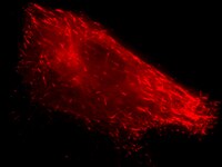
| Application Notes | Fluorescence Microscopy Imaging: REF52 cells were plated in a coverglass chamber slide and transduced with lentiviral particles at an MOI of 20 for 24 hours. After media replacement and 48 hours further incubation, cells were imaged live by oil immersion wide-field fluorescence microscopy. The EB3-RFP displays a punctate distribution characteristic of microtubule ends. Immunocytochemistry Comparison and Inhibitor Analysis: HeLa cells were plated in a coverglass chamber slide and transduced with lentiviral particles at an MOI of 20 for 24 hours. After 24 hours, media was replaced and cells were then further incubated for 48 hours. EB3-RFP displays a nearly complete overlap with EB3 detected by immunocytochemical staining with rabbit polyclonal anti-EB3 (EMD Millipore cat. no. AB6033). Additional Cell Types: MCF7 cells were transduced with EB3-RFP lentivirus as in Figure 1 at an MOI 40 for 24 hours, then stained for tubulin. EB3-RFP displays a punctate and short linear distribution. Immunocytochemical staining with a monoclonal antibody against α-tubulin (EMD Millipore Cat. No. 05-829) reveals the typical filamentous pattern of microtubules. Overlay of EB3-RFP (red), anti-tubulin (green) and DAPI (blue) demonstrates that EB3-RFP is localized to the microtubule ends. Hard-to-transfect Cell Types: Primary cell type HUVEC and Human mesenchymal stem cells (HuMSC) were plated in coverglass chamber slides and transduced with lentiviral particles at an MOI of 20 and 40, respectively, for 24 hours. Time-lapse Imaging: REF52 cells were plated in coverglass chamber slides and transduced with EB3-RFP lentiviral particles. Images were collected every 3 seconds for a total of 2 min. Shown here are 3 sequential frames; to see the entire sequence, please visit our Cell Structure-Biosensors page. For optimal fluorescent visualization, it is recommended to analyze the target expression level within 24-48 hrs after transfection/infection for optimal live cell analysis, as fluorescent intensity may dim over time, especially in difficult-to-transfect cell lines. Infected cells may be frozen down after successful transfection/infection and thawed in culture to retain positive fluorescent expression beyond 24-48 hrs. Length and intensity of fluorescent expression varies between cell lines. Higher MOIs may be required for difficult-to-transfect cell lines. |
| Biological Information | |
|---|---|
| Gene Symbol |
|
| Purification Method | PEG precipitation |
| UniProt Number | |
| Physicochemical Information |
|---|
| Dimensions |
|---|
| Materials Information |
|---|
| Toxicological Information |
|---|
| Safety Information according to GHS |
|---|
| Safety Information |
|---|
| Product Usage Statements | |
|---|---|
| Quality Assurance | Evaluated by transduction of HT-1080 cells and fluorescent imaging performed for assessment of transduction efficiency. |
| Usage Statement |
|
| Packaging Information | |
|---|---|
| Material Size | 1 vial (minimum of 3 x 10E8 IFU/mL) |
| Transport Information |
|---|
| Supplemental Information |
|---|
| Specifications |
|---|
| Global Trade Item Number | |
|---|---|
| Bestellnummer | GTIN |
| 17-10222 | 04053252698040 |
Documentation
LentiBrite™ EB3-RFP Lentiviral Biosensor SDB
| Titel |
|---|


 Antibody[191270-ALL].jpg)
