Wenn Sie das Fenster schließen, wird Ihre Konfiguration nicht gespeichert, es sei denn, Sie haben Ihren Artikel in die Bestellung aufgenommen oder zu Ihren Favoriten hinzugefügt.
Klicken Sie auf OK, um das MILLIPLEX® MAP-Tool zu schließen oder auf Abbrechen, um zu Ihrer Auswahl zurückzukehren.
Wählen Sie konfigurierbare Panels & Premixed-Kits - ODER - Kits für die zelluläre Signaltransduktion & MAPmates™
Konfigurieren Sie Ihre MILLIPLEX® MAP-Kits und lassen sich den Preis anzeigen.
Konfigurierbare Panels & Premixed-Kits
Unser breites Angebot enthält Multiplex-Panels, für die Sie die Analyten auswählen können, die am besten für Ihre Anwendung geeignet sind. Unter einem separaten Register können Sie das Premixed-Cytokin-Format oder ein Singleplex-Kit wählen.
Kits für die zelluläre Signaltransduktion & MAPmates™
Wählen Sie gebrauchsfertige Kits zur Erforschung gesamter Signalwege oder Prozesse. Oder konfigurieren Sie Ihre eigenen Kits mit Singleplex MAPmates™.
Die folgenden MAPmates™ sollten nicht zusammen analysiert werden: -MAPmates™, die einen unterschiedlichen Assaypuffer erfordern. -Phosphospezifische und MAPmate™ Gesamtkombinationen wie Gesamt-GSK3β und Gesamt-GSK3β (Ser 9). -PanTyr und locusspezifische MAPmates™, z.B. Phospho-EGF-Rezeptor und Phospho-STAT1 (Tyr701). -Mehr als 1 Phospho-MAPmate™ für ein einziges Target (Akt, STAT3). -GAPDH und β-Tubulin können nicht mit Kits oder MAPmates™, die panTyr enthalten, analysiert werden.
.
Bestellnummer
Bestellinformationen
St./Pkg.
Liste
Dieser Artikel wurde zu Ihren Favoriten hinzugefügt.
Wählen Sie bitte Spezies, Panelart, Kit oder Probenart
Um Ihr MILLIPLEX® MAP-Kit zu konfigurieren, wählen Sie zunächst eine Spezies, eine Panelart und/oder ein Kit.
Custom Premix Selecting "Custom Premix" option means that all of the beads you have chosen will be premixed in manufacturing before the kit is sent to you.
Catalogue Number
Ordering Description
Qty/Pack
List
Dieser Artikel wurde zu Ihren Favoriten hinzugefügt.
Spezies
Panelart
Gewähltes Kit
Menge
Bestellnummer
Bestellinformationen
St./Pkg.
Listenpreis
96-Well Plate
Menge
Bestellnummer
Bestellinformationen
St./Pkg.
Listenpreis
Weitere Reagenzien hinzufügen (MAPmates erfordern die Verwendung eines Puffer- und Detektionskits)
Menge
Bestellnummer
Bestellinformationen
St./Pkg.
Listenpreis
48-602MAG
Buffer Detection Kit for Magnetic Beads
1 Kit
Platzsparende Option Kunden, die mehrere Kits kaufen, können ihre Multiplex-Assaykomponenten in Kunststoffbeuteln anstelle von Packungen erhalten, um eine kompaktere Lagerung zu ermöglichen.
Dieser Artikel wurde zu Ihren Favoriten hinzugefügt.
Das Produkt wurde in Ihre Bestellung aufgenommen
Sie können nun ein weiteres Kit konfigurieren, ein Premixed-Kit wählen, zur Kasse gehen oder das Bestell-Tool schließen.
Use Anti-TLS Antibody, clone 1FU-1D2 (Mouse Monoclonal Antibody) validated in WB, ICC to detect TLS also known as 75 kDa DNA-pairing protein, FUS-CHOP fusion protein.
More>>Use Anti-TLS Antibody, clone 1FU-1D2 (Mouse Monoclonal Antibody) validated in WB, ICC to detect TLS also known as 75 kDa DNA-pairing protein, FUS-CHOP fusion protein. Less<<
Anti-TLS Antibody, clone 1FU-1D2: SDB (Sicherheitsdatenblätter), Analysenzertifikate und Qualitätszertifikate, Dossiers, Broschüren und andere verfügbare Dokumente.
The TLS/FUS protein contains an RNA-recognition motif and is a component of nuclear riboprotein complexes. It resembles a transcription factor in that it binds DNA, contributes a transcriptional activation domain to the FUS-ERG oncoprotein, and interacts with several transcription factors in vitro. It binds both single-stranded and double-stranded DNA and promotes ATP-independent annealing of complementary single-stranded DNAs and D-loop formation in superhelical double-stranded DNA. It may play a role in maintenance of genomic integrity.
Use Anti-TLS Antibody, clone 1FU-1D2 (Mouse Monoclonal Antibody) validated in WB, ICC to detect TLS also known as 75 kDa DNA-pairing protein, FUS-CHOP fusion protein.
Key Applications
Western Blotting
Immunocytochemistry
Application Notes
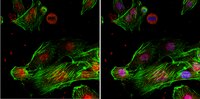
Immunocytochemistry Analysis: 1:500 dilution from a representative lot detected TLS in C2C12 cells.
This gene encodes a multifunctional protein component of the heterogeneous nuclear ribonucleoprotein (hnRNP) complex. The hnRNP complex is involved in pre-mRNA splicing and the export of fully processed mRNA to the cytoplasm. This protein belongs to the FET family of RNA-binding proteins which have been implicated in cellular processes that include regulation of gene expression, maintenance of genomic integrity and mRNA/microRNA processing. Alternative splicing results in multiple transcript variants. Defects in this gene result in amyotrophic lateral sclerosis type 6. [provided by RefSeq].
FUNCTION: Binds both single-stranded and double-stranded DNA and promotes ATP-independent annealing of complementary single-stranded DNAs and D-loop formation in superhelical double-stranded DNA. May play a role in maintenance of genomic integrity.
SUBUNIT STRUCTURE: Component of nuclear riboprotein complexes. Interacts with ILF3, TDRD3 and SF1. Interacts through its C-terminus with SFRS13A. Interacts with OTUB1 and SARNP.
SUBCELLULAR LOCATION: Nucleus
TISSUE SPECIFICITY: Ubiquitous.
PTM: Arg-216 and Arg-218 are dimethylated, probably to asymmetric dimethylarginine.
INVOLVEMENT IN DISEASE: A chromosomal aberration involving FUS is a cause of a form of malignant myxoid liposarcoma. Translocation t(12;16)(q13;p11) with DDIT3.
A chromosomal aberration involving FUS is a cause of acute myeloid leukemia (AML). Translocation t(16;21)(p11;q22) with ERG.
A chromosomal aberration involving FUS is associated with angiomatoid fibrous histiocytoma (AFH) [MIM:612160]. Translocation t(12;16)(q13;p11.2) with ATF1 generates a chimeric FUS/ATF1 protein.
Defects in FUS are the cause of amyotrophic lateral sclerosis type 6 (ALS6) [MIM:608030]. ALS6 is a familial form of amyotrophic lateral sclerosis. ALS is a neurodegenerative disorder affecting upper motor neurons in the brain and lower motor neurons in the brain stem and spinal cord, resulting in fatal paralysis. Sensory abnormalities are absent. Death usually occurs within 2 to 5 years. The etiology of amyotrophic lateral sclerosis is likely to be multifactorial, involving both genetic and environmental factors. The disease is inherited in 5-10%.
SEQUENCE SIMILARITIES: Belongs to the RRM TET family.
Contains 1 RanBP2-type zinc finger.
Contains 1 RRM (RNA recognition motif) domain.
Molecular Weight
Observed at 65 kDa
Physicochemical Information
Dimensions
Materials Information
Toxicological Information
Safety Information according to GHS
Safety Information
Product Usage Statements
Quality Assurance
Evaluated by Western Blot in HepG2 cell lysate.
Western Blot Analysis: 1:1,000 dilution of this antibody detected TLS on 10 µg of HepG2 cell lysate.
Usage Statement
Unless otherwise stated in our catalog or other company documentation accompanying the product(s), our products are intended for research use only and are not to be used for any other purpose, which includes but is not limited to, unauthorized commercial uses, in vitro diagnostic uses, ex vivo or in vivo therapeutic uses or any type of consumption or application to humans or animals.
Storage and Shipping Information
Storage Conditions
Stable for 1 year at -20°C from date of receipt. Handling Recommendations: Upon receipt and prior to removing the cap, centrifuge the vial and gently mix the solution. Aliquot into microcentrifuge tubes and store at -20°C. Avoid repeated freeze/thaw cycles, which may damage IgG and affect product performance.