The transforming acidic coiled coil proteins interact with nuclear histone acetyltransferases.
Gangisetty, O; Lauffart, B; Sondarva, GV; Chelsea, DM; Still, IH
Oncogene
23
2559-63
2004
Abstract anzeigen
Dysregulation of the human transforming acidic coiled coil (TACC) genes is thought to be important in the development of multiple myeloma, breast and gastric cancer. However, even though these proteins have been implicated in the control of cell growth and differentiation, the mechanism by which they function still remains to be clarified. Using the yeast two-hybrid assay, we have now identified the histone acetyltransferase (HAT) hGCN5L2 as a TACC2-binding protein. GST pull-down analysis subsequently confirmed that all human TACC family members can bind in vitro to hGCN5L2. The authenticity of these interactions was validated by coimmunoprecipitation assays within the human embryonic kidney cell line HEK293, which identified the TACC2s isoform as a component consistently bound to several different members of HAT family. This raises the possibility that aberrant expression of one or more TACC proteins may affect gene regulation through their interaction with components of chromatin remodeling complexes, thus contributing to tumorigenesis. | | | 14767476
 |
Molecular cloning, genomic structure and interactions of the putative breast tumor suppressor TACC2.
Brenda Lauffart, Omkaram Gangisetty, Ivan H Still
Genomics
81
192-201
2003
Abstract anzeigen
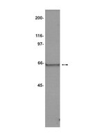
The human transforming acidic coiled-coil 2 (TACC2) gene has been suggested recently to be a putative breast tumor suppressor. Now we can report the cloning of full length TACC2 cDNAs corresponding to the major isoforms expressed during development. The TACC2 gene is encoded by 23 exons, and spans 255 kb of chromosome 10q26. In breast cancer cell lines, TACC2 is expressed as a 120 kDa protein corresponding to the major transcript expressed in the mammary gland. Although only slight differences in the expression of TACC2 in normal versus breast tumors were observed, overexpression of TACC2 can alter the in vitro cellular dynamics of some breast cancer cell lines. Significantly, we demonstrate that TACC2 interacts with GAS41 and the SWI/SNF chromatin remodeling complex. This suggests that defects in TACC2 expression may affect gene regulation, thus contributing to the pathogenesis of some tumors. | | | 12620397
 |
TACC1-chTOG-Aurora A protein complex in breast cancer.
Conte, N; Delaval, B; Ginestier, C; Ferrand, A; Isnardon, D; Larroque, C; Prigent, C; Séraphin, B; Jacquemier, J; Birnbaum, D
Oncogene
22
8102-16
2003
Abstract anzeigen
The three human TACC (transforming acidic coiled-coil) genes encode a family of proteins with poorly defined functions that are suspected to play a role in oncogenesis. A Xenopus TACC homolog called Maskin is involved in translational control, while Drosophila D-TACC interacts with the microtubule-associated protein MSPS (Mini SPindleS) to ensure proper dynamics of spindle pole microtubules during cell division. We have delineated here the interactions of TACC1 with four proteins, namely the microtubule-associated chTOG (colonic and hepatic tumor-overexpressed gene) protein (ortholog of Drosophila MSPS), the adaptor protein TRAP (tudor repeat associator with PCTAIRE2), the mitotic serine/threonine kinase Aurora A and the mRNA regulator LSM7 (Like-Sm protein 7). To measure the relevance of the TACC1-associated complex in human cancer we have examined the expression of the three TACC, chTOG and Aurora A in breast cancer using immunohistochemistry on tissue microarrays. We show that expressions of TACC1, TACC2, TACC3 and Aurora A are significantly correlated and downregulated in a subset of breast tumors. Using siRNAs, we further show that depletion of chTOG and, to a lesser extent of TACC1, perturbates cell division. We propose that TACC proteins, which we also named 'Taxins', control mRNA translation and cell division in conjunction with microtubule organization and in association with chTOG and Aurora A, and that these complexes and cell processes may be affected during mammary gland oncogenesis. | | | 14603251
 |
Distinct and complementary information provided by use of tissue and DNA microarrays in the study of breast tumor markers.
Ginestier, C; Charafe-Jauffret, E; Bertucci, F; Eisinger, F; Geneix, J; Bechlian, D; Conte, N; Adélaïde, J; Toiron, Y; Nguyen, C; Viens, P; Mozziconacci, MJ; Houlgatte, R; Birnbaum, D; Jacquemier, J
The American journal of pathology
161
1223-33
2002
Abstract anzeigen
Emerging high-throughput screening technologies are rapidly providing opportunities to identify new diagnostic and prognostic markers and new therapeutic targets in human cancer. Currently, cDNA arrays allow the quantitative measurement of thousands of mRNA expression levels simultaneously. Validation of this tool in hospital settings can be done on large series of archival paraffin-embedded tumor samples using the new technique of tissue microarray. On a series of 55 clinically and pathologically homogeneous breast tumors, we compared for 15 molecules with a proven or suspected role in breast cancer, the mRNA expression levels measured by cDNA array analysis with protein expression levels obtained using tumor tissue microarrays. The validity of cDNA array and tissue microarray data were first verified by comparison with quantitative reverse transcriptase-polymerase chain reaction measurements and immunohistochemistry on full tissue sections, respectively. We found a good correlation between cDNA and tissue array analyses in one-third of the 15 molecules, and no correlation in the remaining two-thirds. Furthermore, protein but not RNA levels may have prognostic value; this was the case for MUC1 protein, which was studied further using a tissue microarray containing approximately 600 tumor samples. For THBS1 the opposite was observed because only RNA levels had prognostic value. Thus, differences extended to clinical prognostic information obtained by the two methods underlining their complementarity and the need for a global molecular analysis of tumors at both the RNA and protein levels. | Immunohistochemistry | Human | 12368196
 |


