Wenn Sie das Fenster schließen, wird Ihre Konfiguration nicht gespeichert, es sei denn, Sie haben Ihren Artikel in die Bestellung aufgenommen oder zu Ihren Favoriten hinzugefügt.
Klicken Sie auf OK, um das MILLIPLEX® MAP-Tool zu schließen oder auf Abbrechen, um zu Ihrer Auswahl zurückzukehren.
Wählen Sie konfigurierbare Panels & Premixed-Kits - ODER - Kits für die zelluläre Signaltransduktion & MAPmates™
Konfigurieren Sie Ihre MILLIPLEX® MAP-Kits und lassen sich den Preis anzeigen.
Konfigurierbare Panels & Premixed-Kits
Unser breites Angebot enthält Multiplex-Panels, für die Sie die Analyten auswählen können, die am besten für Ihre Anwendung geeignet sind. Unter einem separaten Register können Sie das Premixed-Cytokin-Format oder ein Singleplex-Kit wählen.
Kits für die zelluläre Signaltransduktion & MAPmates™
Wählen Sie gebrauchsfertige Kits zur Erforschung gesamter Signalwege oder Prozesse. Oder konfigurieren Sie Ihre eigenen Kits mit Singleplex MAPmates™.
Die folgenden MAPmates™ sollten nicht zusammen analysiert werden: -MAPmates™, die einen unterschiedlichen Assaypuffer erfordern. -Phosphospezifische und MAPmate™ Gesamtkombinationen wie Gesamt-GSK3β und Gesamt-GSK3β (Ser 9). -PanTyr und locusspezifische MAPmates™, z.B. Phospho-EGF-Rezeptor und Phospho-STAT1 (Tyr701). -Mehr als 1 Phospho-MAPmate™ für ein einziges Target (Akt, STAT3). -GAPDH und β-Tubulin können nicht mit Kits oder MAPmates™, die panTyr enthalten, analysiert werden.
.
Bestellnummer
Bestellinformationen
St./Pkg.
Liste
Dieser Artikel wurde zu Ihren Favoriten hinzugefügt.
Wählen Sie bitte Spezies, Panelart, Kit oder Probenart
Um Ihr MILLIPLEX® MAP-Kit zu konfigurieren, wählen Sie zunächst eine Spezies, eine Panelart und/oder ein Kit.
Custom Premix Selecting "Custom Premix" option means that all of the beads you have chosen will be premixed in manufacturing before the kit is sent to you.
Catalogue Number
Ordering Description
Qty/Pack
List
Dieser Artikel wurde zu Ihren Favoriten hinzugefügt.
Spezies
Panelart
Gewähltes Kit
Menge
Bestellnummer
Bestellinformationen
St./Pkg.
Listenpreis
96-Well Plate
Menge
Bestellnummer
Bestellinformationen
St./Pkg.
Listenpreis
Weitere Reagenzien hinzufügen (MAPmates erfordern die Verwendung eines Puffer- und Detektionskits)
Menge
Bestellnummer
Bestellinformationen
St./Pkg.
Listenpreis
48-602MAG
Buffer Detection Kit for Magnetic Beads
1 Kit
Platzsparende Option Kunden, die mehrere Kits kaufen, können ihre Multiplex-Assaykomponenten in Kunststoffbeuteln anstelle von Packungen erhalten, um eine kompaktere Lagerung zu ermöglichen.
Dieser Artikel wurde zu Ihren Favoriten hinzugefügt.
Das Produkt wurde in Ihre Bestellung aufgenommen
Sie können nun ein weiteres Kit konfigurieren, ein Premixed-Kit wählen, zur Kasse gehen oder das Bestell-Tool schließen.
ABN1714
Sigma-AldrichAnti-Rootletin
Anti-Rootletin Antibody, Cat. No. ABN1714, is a highly specific rabbit polyclonal antibody that targets Rootletin and has been tested in Immunocytochemistry, Immunohistochemistry (Paraffin), and Immunoprecipitation.
More>>Anti-Rootletin Antibody, Cat. No. ABN1714, is a highly specific rabbit polyclonal antibody that targets Rootletin and has been tested in Immunocytochemistry, Immunohistochemistry (Paraffin), and Immunoprecipitation. Less<<
Anti-Rootletin: SDB (Sicherheitsdatenblätter), Analysenzertifikate und Qualitätszertifikate, Dossiers, Broschüren und andere verfügbare Dokumente.
Rootletin (UniProt: Q8CJ40; also known as Ciliary rootlet coiled-coil protein) is encoded by the Crocc (also known as Kiaa0445) gene (Gene ID: 230872) in murine species. Rootletin is a major structural component of the ciliary rootlet, a cytoskeletal-like structure in ciliated cells that originates from the basal body at the proximal end of a cilium and extends proximally toward the cell nucleus. It contributes to centrosome cohesion before mitosis. In ciliated cells, it is associated with ciliary rootlets, but in non-ciliated cells it is localized between, around and at the proximal ends of the centrioles. It dissociates from the centrioles at the onset of D7mitosis and re-associates with them at anaphase. Highest expression of Rootletin is found in photoreceptor cells of retina. Lower expressions have been reported in brain, trachea and kidney. In retinal photoreceptors rootlets extend from the basal bodies to the synaptic terminals and anchor endoplasmic reticulum membranes along their length. During embryonic development, it is enriched along the apical domains of neuro epithelium in brain ventricular zone, in primordia of retinal pigment epithelia, and in neural retina. Rootletin contains a globular head domain and a tail domain that consists of extended coiled-coil structures. The head domain may be required for targeting to the basal body and binding to a kinesin light chain. (Ref.: Yang, J., et al. (2002). J. Cell Biol. 159(3): 431-440).
References
Product Information
Format
Affinity Purified
Presentation
Purified rabbit polyclonal antibody in PBS with 50% glycerol and without azide.
Anti-Rootletin Antibody, Cat. No. ABN1714, is a highly specific rabbit polyclonal antibody that targets Rootletin and has been tested in Immunocytochemistry, Immunohistochemistry (Paraffin), and Immunoprecipitation.
Key Applications
Immunoprecipitation
Immunohistochemistry (Paraffin)
Immunocytochemistry
Application Notes
Immunohistochemistry Analysis: A 1:50-250 dilution from a representative lot detected Rootletin in mouse cerebral cortex and mouse retina tissues.
Immunocytochemistry Analysis: A representative lot detected Rootletin in Immunocytochemistry applications (Veleri, S., et. al. (2014). Nat Commun. 5:4207).
Immunoprecipitation Analysis: A representative lot detected Rootletin in Immunoprecipitation applications (Yang, J., et. al. (2002). J Cell Biol. 159(3):431-40).
Immunofluorescence Analysis: A representative lot detected Rootletin in Immunofluorescence applications (Sun, X., et. al. (2012). Cilia. 1(1):21).
Biological Information
Immunogen
His-tagged recombinant fragment corresponding to 188 amino acids from the C-terminal region of murine Rootletin.
Concentration
Please refer to lot specific datasheet.
Host
Rabbit
Specificity
This rabbit polyclonal antibody detects Rootletin in murine hippocampus and cerebral cortex. It targets an epitope within 188 amino acids from the C-terminal region.
~227 kDa observed; 226.95 kDa calculated. Uncharacterized bands may be observed in some lysate(s).
Physicochemical Information
Dimensions
Materials Information
Toxicological Information
Safety Information according to GHS
Safety Information
Product Usage Statements
Quality Assurance
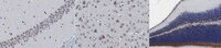
Evaluated by Immunohistochemistry in mouse hippocampus.
Immunohistochemistry Analysis: A 1:50 dilution of this antibody detected Rootletin in mouse hippocampus.
Usage Statement
Unless otherwise stated in our catalog or other company documentation accompanying the product(s), our products are intended for research use only and are not to be used for any other purpose, which includes but is not limited to, unauthorized commercial uses, in vitro diagnostic uses, ex vivo or in vivo therapeutic uses or any type of consumption or application to humans or animals.
Storage and Shipping Information
Storage Conditions
Stable for 1 year at -20°C from date of receipt. Handling Recommendations: Upon receipt and prior to removing the cap, centrifuge the vial and gently mix the solution. Aliquot into microcentrifuge tubes and store at -20°C. Avoid repeated freeze/thaw cycles, which may damage IgG and affect product performance.