Wenn Sie das Fenster schließen, wird Ihre Konfiguration nicht gespeichert, es sei denn, Sie haben Ihren Artikel in die Bestellung aufgenommen oder zu Ihren Favoriten hinzugefügt.
Klicken Sie auf OK, um das MILLIPLEX® MAP-Tool zu schließen oder auf Abbrechen, um zu Ihrer Auswahl zurückzukehren.
Wählen Sie konfigurierbare Panels & Premixed-Kits - ODER - Kits für die zelluläre Signaltransduktion & MAPmates™
Konfigurieren Sie Ihre MILLIPLEX® MAP-Kits und lassen sich den Preis anzeigen.
Konfigurierbare Panels & Premixed-Kits
Unser breites Angebot enthält Multiplex-Panels, für die Sie die Analyten auswählen können, die am besten für Ihre Anwendung geeignet sind. Unter einem separaten Register können Sie das Premixed-Cytokin-Format oder ein Singleplex-Kit wählen.
Kits für die zelluläre Signaltransduktion & MAPmates™
Wählen Sie gebrauchsfertige Kits zur Erforschung gesamter Signalwege oder Prozesse. Oder konfigurieren Sie Ihre eigenen Kits mit Singleplex MAPmates™.
Die folgenden MAPmates™ sollten nicht zusammen analysiert werden: -MAPmates™, die einen unterschiedlichen Assaypuffer erfordern. -Phosphospezifische und MAPmate™ Gesamtkombinationen wie Gesamt-GSK3β und Gesamt-GSK3β (Ser 9). -PanTyr und locusspezifische MAPmates™, z.B. Phospho-EGF-Rezeptor und Phospho-STAT1 (Tyr701). -Mehr als 1 Phospho-MAPmate™ für ein einziges Target (Akt, STAT3). -GAPDH und β-Tubulin können nicht mit Kits oder MAPmates™, die panTyr enthalten, analysiert werden.
.
Bestellnummer
Bestellinformationen
St./Pkg.
Liste
Dieser Artikel wurde zu Ihren Favoriten hinzugefügt.
Wählen Sie bitte Spezies, Panelart, Kit oder Probenart
Um Ihr MILLIPLEX® MAP-Kit zu konfigurieren, wählen Sie zunächst eine Spezies, eine Panelart und/oder ein Kit.
Custom Premix Selecting "Custom Premix" option means that all of the beads you have chosen will be premixed in manufacturing before the kit is sent to you.
Catalogue Number
Ordering Description
Qty/Pack
List
Dieser Artikel wurde zu Ihren Favoriten hinzugefügt.
Spezies
Panelart
Gewähltes Kit
Menge
Bestellnummer
Bestellinformationen
St./Pkg.
Listenpreis
96-Well Plate
Menge
Bestellnummer
Bestellinformationen
St./Pkg.
Listenpreis
Weitere Reagenzien hinzufügen (MAPmates erfordern die Verwendung eines Puffer- und Detektionskits)
Menge
Bestellnummer
Bestellinformationen
St./Pkg.
Listenpreis
48-602MAG
Buffer Detection Kit for Magnetic Beads
1 Kit
Platzsparende Option Kunden, die mehrere Kits kaufen, können ihre Multiplex-Assaykomponenten in Kunststoffbeuteln anstelle von Packungen erhalten, um eine kompaktere Lagerung zu ermöglichen.
Dieser Artikel wurde zu Ihren Favoriten hinzugefügt.
Das Produkt wurde in Ihre Bestellung aufgenommen
Sie können nun ein weiteres Kit konfigurieren, ein Premixed-Kit wählen, zur Kasse gehen oder das Bestell-Tool schließen.
Proteinase-activated receptor 4 (UniProt Q96RI0; also known as Coagulation factor II receptor-like 3, PAR-4, Thrombin receptor-like 3) is encoded by the F2RL3 (also known as PAR4) gene (Gene ID 9002) in human. Protease-activated receptors (PARs) constitute a unique family of seven-transmembrane, G-protein-coupled receptors (GPCRs) activated by proteolytic cleavage of their N-terminal propeptide sequence. Once cleaved off, the N-terminal propeptide fragment functions as a ligand and activates the receptor by binding the second extracellular loop. The four PAR family members (PAR-1 to PAR-4) are widely expressed and activated by multiple proteases, and utilize different types of G-proteins (Gi, Gq, and G12/13) for signal transdution depending on the activating protease and cellular context. PAR-4 is expressed on platelets and exhibits a low-affinity for thrombin. However, PAR-4 is able to form hetero-oligomers with both PAR-1 and the ADP receptor P2Y12 to mediate thrombin- and ADP-initiated signaling. PAR-4 cleavage is significantly enhanced through hetero-oligomerization with PAR-1, and PAR-4 interaction with P2Y12 is directly linked to arrestin-2 recruitment and AKT signaling. PAR-4 is a 7-transmembrane (a.a. 83-103, 109-129, 152-172, 192-213, 248-268, 284-304, 320-343) GPCR activated by thrombin cleavage between R47 and G48, having 3 extracellular loops and 3 intracellular loops between the extracellular N-terminal end (a.a. 48-82) the cytoplasmic C-terminal tail (a.a. 344-385).
References
Product Information
Format
Purified
Presentation
Purified mouse monoclonal IgG2bλ antibody in PBS without preservatives.
Anti-PAR4 Antibody, clone 5F10 is an antibody against PAR4 for use in Western Blotting, Immunocytochemistry, Flow Cytometry, Inhibition.
Key Applications
Western Blotting
Immunocytochemistry
Flow Cytometry
Inhibition
Application Notes
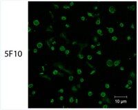
Immunocytochemistry Analysis: 20 µg/mL from a representative lot detected endogenous PAR4 by fluorescent immunocytochemistry staining of freshly isolated human platelets (Courtesy of Dr. Marvin Nieman, Case Western Reserve University, Cleveland, OH, USA). Immunocytochemistry Analysis: A representative lot detected the expression of exogenously transfected human PAR4 by fluorescent immunocytochemistry staining of 4% formaldehyde-fixed HEK293 Flp-In cells following tetracycline treatment (Mumaw, M.M., et al. (2015). Thromb. Res. 135(6):1165-1171). Flow Cytometry Analysis: A representative lot detected tetracycline-induced expression of exogenously transfected human PAR4 on the surface of HEK293 Flp-In cells. Thrombin treatment diminished cell surface PAR4 immunoreactivity (Mumaw, M.M., et al. (2015). Thromb. Res. 135(6):1165-1171). Inhibition Analysis: A representative lot, when added prior to thrombin, protected cell surface PAR4 against thrombin cleavage (Mumaw, M.M., et al. (2015). Thromb. Res. 135(6):1165-1171). Western Blotting Analysis: A representative lot detected MBP fusion proteins containing human PAR4 fragment a.a. 18-78 or 41-66, but not 48-72. MBP-PAR4 fusion degradation by thrombin treatment ablolished target band detection by clone 5F10 (Mumaw, M.M., et al. (2015). Thromb. Res. 135(6):1165-1171). Western Blotting Analysis: A representative lot detected tetracycline-induced expression of exogenously introduced human PAR4 in a HEK293 Flp-In cell line, as well as endogenous PAR4 in isolated human platelets (hPLTs). Thrombin activation of hPLTs diminished PAR4 target band detection (Mumaw, M.M., et al. (2015). Thromb. Res. 135(6):1165-1171).
Biological Information
Immunogen
MBP-conjugated recombinant human PAR4 N-terminal fragment including the propeptide sequence.
Epitope
Near (N-terminal to) the thrombin cleavage site.
Clone
5F10
Concentration
Please refer to lot specific datasheet.
Host
Mouse
Specificity
Clone 5F10 recognizes a propeptide epitope near (N-terminal to) the thrombin-cleavage site and protects cell surface PAR4 against thrombin cleavage (Mumaw, M.M., et al. (2015). Thromb. Res. 135(6):1165-1171). Clone 5F10 recognizes prepro- and pro-, but not proteolytically activated, forms of human PAR4.
~45 kDa observed. 41.13/39.16 kDa (prepro-/pro-PAR4) calculated. The broad banding pattern and larger apparent band size is consistent with the detection of glycosylated PAR4. Uncharacterized band(s) may appear in some lysates.
Physicochemical Information
Dimensions
Materials Information
Toxicological Information
Safety Information according to GHS
Safety Information
Product Usage Statements
Quality Assurance
Evaluated by Western Blotting in human platelet lysate.
Western Blotting Analysis: A 1:250 dilution of this antibody detected PAR4 in 50 µg of human platelet lysate.
Usage Statement
Unless otherwise stated in our catalog or other company documentation accompanying the product(s), our products are intended for research use only and are not to be used for any other purpose, which includes but is not limited to, unauthorized commercial uses, in vitro diagnostic uses, ex vivo or in vivo therapeutic uses or any type of consumption or application to humans or animals.