


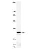
MAB312RX Sigma-AldrichAnti-Neuron Cell Surface Antigen Antibody, clone A2B5-105 (7), Alexa Fluor® 488


Anti-Neuron Cell Surface Antigen Antibody, clone A2B5-105 (7), Alexa Fluor 488 detects level of Neuron Cell Surface Antigen & has been published & validated for use in IC, IH.
More>> Anti-Neuron Cell Surface Antigen Antibody, clone A2B5-105 (7), Alexa Fluor 488 detects level of Neuron Cell Surface Antigen & has been published & validated for use in IC, IH. Less<<SDB (Sicherheitsdatenblätter), Analysenzertifikate und Qualitätszertifikate, Dossiers, Broschüren und andere verfügbare Dokumente.
Empfohlene Produkte


Übersicht
| Replacement Information |
|---|
Key Spec Table
| Species Reactivity | Key Applications | Host | Format | Antibody Type |
|---|---|---|---|---|
| Ch, H, M, R | ICC, IHC | M | AlexaFluor®488 | Monoclonal Antibody |
| Description | |
|---|---|
| Catalogue Number | MAB312RX |
| Brand Family | Chemicon® |
| Trade Name |
|
| Description | Anti-Neuron Cell Surface Antigen Antibody, clone A2B5-105 (7), Alexa Fluor® 488 |
| Alternate Names |
|
| References |
|---|
| Product Information | |
|---|---|
| Format | AlexaFluor®488 |
| HS Code | 3002 15 90 |
| Presentation | Purified immunoglobulin conjugated to Alexa Fluor® 488. Liquid in Phosphate buffer with 15 mg/mL BSA as a stabilizer and 0.1% sodium azide. |
| Quality Level | MQ100 |
| Physicochemical Information |
|---|
| Dimensions |
|---|
| Materials Information |
|---|
| Toxicological Information |
|---|
| Safety Information according to GHS |
|---|
| Safety Information |
|---|
| Storage and Shipping Information | |
|---|---|
| Storage Conditions | Maintain at 2-8°C in undiluted aliquots in the dark for up to 6 months after date of receipt. |
| Packaging Information | |
|---|---|
| Material Size | 100 µg |
| Transport Information |
|---|
| Supplemental Information |
|---|
| Specifications |
|---|
| Global Trade Item Number | |
|---|---|
| Bestellnummer | GTIN |
| MAB312RX | 04053252318115 |
Documentation
Anti-Neuron Cell Surface Antigen Antibody, clone A2B5-105 (7), Alexa Fluor® 488 SDB
| Titel |
|---|
Anti-Neuron Cell Surface Antigen Antibody, clone A2B5-105 (7), Alexa Fluor® 488 Analysenzertifikate
Literatur
| Übersicht | Pub Med ID |
|---|---|
| High-mobility Group Box-1 and Its Receptors Contribute to Proinflammatory Response in the Acute Phase of Spinal Cord Injury in Rats. Chen KB, Uchida K, Nakajima H, Yayama T, Hirai T, Guerrero AR, Kobayashi S, Ma WY, Liu SY, Zhu P, Baba H Spine (Phila Pa 1976) 2010 Abstract anzeigen | 21343866
|
| The effect of TGF-beta2 on elastin, type VI collagen, and components of the proteolytic degradation system in human optic nerve astrocytes. Neumann, Carolin, et al. Invest. Ophthalmol. Vis. Sci., 49: 1464-72 (2008) 2008 | 18385064
|
| Pancreatic islet A2B5- and 3G5-reactive gangliosides are markers of differentiation in rat insulinoma cells. Bartholomeusz, R K, et al. Endocrinology, 124: 2680-5 (1989) 1988 Abstract anzeigen | 2541995
|
| Flow cytometric analysis of rat striatal nerve terminals Wolf, M E and Kapatos, G J Neurosci, 9:94-105 (1989) 1988 | 2563283
|
| Monoclonal antibody to embryonic CNS antigen A2B5 provides evidence for the involvement of membrane components at sites of Alzheimer degeneration and detects sulfatides as well as gangliosides. Majocha, R E, et al. J. Neurochem., 53: 953-61 (1989) 1988 | 2668446
|
| Schwann cells influence the expression of ganglioside GD3 by rat dorsal root ganglion neurons. Levison, S W and McCarthy, K D J. Neuroimmunol., 24: 223-32 (1989) 1988 Abstract anzeigen | 2681262
|
| Lymphokines facilitate maturation of oligodendrocytes in vitro. Suzumura, A and Silberberg, D H Brain Res., 480: 51-7 (1989) 1988 | 2713667
|
| Bone marrow examination in neuroblastoma patients: a morphologic, immunocytochemical, and immunohistochemical study Oppedal, B R, et al Hum Pathol, 20:800-805 (1989) 1988 | 2744752
|
| CNS neuronal cell line-derived factors regulate gliogenesis in neonatal rat brain cultures Bottenstein, J E, et al J Neurosci Res, 20:291-303 (1988) 1987 | 2852260
|
| Immunohistological diagnosis of central nervous system tumours using a monoclonal antibody panel Coakham, H B, et al J Clin Pathol, 38:165-173 (1985) 1985 | 3881481
|