Wenn Sie das Fenster schließen, wird Ihre Konfiguration nicht gespeichert, es sei denn, Sie haben Ihren Artikel in die Bestellung aufgenommen oder zu Ihren Favoriten hinzugefügt.
Klicken Sie auf OK, um das MILLIPLEX® MAP-Tool zu schließen oder auf Abbrechen, um zu Ihrer Auswahl zurückzukehren.
Wählen Sie konfigurierbare Panels & Premixed-Kits - ODER - Kits für die zelluläre Signaltransduktion & MAPmates™
Konfigurieren Sie Ihre MILLIPLEX® MAP-Kits und lassen sich den Preis anzeigen.
Konfigurierbare Panels & Premixed-Kits
Unser breites Angebot enthält Multiplex-Panels, für die Sie die Analyten auswählen können, die am besten für Ihre Anwendung geeignet sind. Unter einem separaten Register können Sie das Premixed-Cytokin-Format oder ein Singleplex-Kit wählen.
Kits für die zelluläre Signaltransduktion & MAPmates™
Wählen Sie gebrauchsfertige Kits zur Erforschung gesamter Signalwege oder Prozesse. Oder konfigurieren Sie Ihre eigenen Kits mit Singleplex MAPmates™.
Die folgenden MAPmates™ sollten nicht zusammen analysiert werden: -MAPmates™, die einen unterschiedlichen Assaypuffer erfordern. -Phosphospezifische und MAPmate™ Gesamtkombinationen wie Gesamt-GSK3β und Gesamt-GSK3β (Ser 9). -PanTyr und locusspezifische MAPmates™, z.B. Phospho-EGF-Rezeptor und Phospho-STAT1 (Tyr701). -Mehr als 1 Phospho-MAPmate™ für ein einziges Target (Akt, STAT3). -GAPDH und β-Tubulin können nicht mit Kits oder MAPmates™, die panTyr enthalten, analysiert werden.
.
Bestellnummer
Bestellinformationen
St./Pkg.
Liste
Dieser Artikel wurde zu Ihren Favoriten hinzugefügt.
Wählen Sie bitte Spezies, Panelart, Kit oder Probenart
Um Ihr MILLIPLEX® MAP-Kit zu konfigurieren, wählen Sie zunächst eine Spezies, eine Panelart und/oder ein Kit.
Custom Premix Selecting "Custom Premix" option means that all of the beads you have chosen will be premixed in manufacturing before the kit is sent to you.
Catalogue Number
Ordering Description
Qty/Pack
List
Dieser Artikel wurde zu Ihren Favoriten hinzugefügt.
Spezies
Panelart
Gewähltes Kit
Menge
Bestellnummer
Bestellinformationen
St./Pkg.
Listenpreis
96-Well Plate
Menge
Bestellnummer
Bestellinformationen
St./Pkg.
Listenpreis
Weitere Reagenzien hinzufügen (MAPmates erfordern die Verwendung eines Puffer- und Detektionskits)
Menge
Bestellnummer
Bestellinformationen
St./Pkg.
Listenpreis
48-602MAG
Buffer Detection Kit for Magnetic Beads
1 Kit
Platzsparende Option Kunden, die mehrere Kits kaufen, können ihre Multiplex-Assaykomponenten in Kunststoffbeuteln anstelle von Packungen erhalten, um eine kompaktere Lagerung zu ermöglichen.
Dieser Artikel wurde zu Ihren Favoriten hinzugefügt.
Das Produkt wurde in Ihre Bestellung aufgenommen
Sie können nun ein weiteres Kit konfigurieren, ein Premixed-Kit wählen, zur Kasse gehen oder das Bestell-Tool schließen.
Anti-MAdCAM-1, clone MECA-367, Cat. No. MABF2072, is a rat monoclonal antibody that detects murine Mucosal addressin cell adhesion molecule 1 (MAdCAM-1) and has been tested for use in Immunohistochemistry (Paraffin), Inhibition/Function assays, Neutralizing, and Western Blotting.
More>>Anti-MAdCAM-1, clone MECA-367, Cat. No. MABF2072, is a rat monoclonal antibody that detects murine Mucosal addressin cell adhesion molecule 1 (MAdCAM-1) and has been tested for use in Immunohistochemistry (Paraffin), Inhibition/Function assays, Neutralizing, and Western Blotting. Less<<
Anti-MAdCAM-1 Antibody, clone MECA-367: SDB (Sicherheitsdatenblätter), Analysenzertifikate und Qualitätszertifikate, Dossiers, Broschüren und andere verfügbare Dokumente.
Mucosal addressin cell adhesion molecule 1 (UniProt: Q61826; also known as MAdCAM-1, mMAdCAM-1) is encoded by the Madcam1 gene (Gene ID: 17123) in murine species. MAdCAM-1 is a cell adhesion leukocyte receptor that helps to direct lymphocyte traffic into mucosal tissues including the Peyer patches and the intestinal lamina propria. It is highly expressed on high endothelial venules (HEV) of organized intestinal lymphoid tissues and mesenteric lymph nodes, and in the lamina propria of the intestine. Its expression is also observed in brain endothelioma cells and mucosal tissues during chronic inflammation. Aberrant expression of MAdCAM-1 has also been reported in the inflamed pancreas. MAdCAM-1 can bind both the integrin alpha-4/beta-7 and L-selectin and is involved in regulating both the passage and retention of leukocytes. It is synthesized with a signal peptide (aa 1-21), which is subsequently cleaved off in mature form. MAdCAM-1 has an extracellular domain (aa 22-109), a transmembrane domain (aa 365-385), and a cytoplasmic domain (aa 386-405). It also contains three Ig-like domains (aa 22-109; 110-227; 258-357) and a Mucin-like region (aa 221-257). Two isoforms of MAdCAM-1 have been described that are produced by alternative splicing. Both isoforms can adhere to integrin alpha-4/beta-7. Isoform 2 that lacks the mucin-like domain is believed to be specialized in supporting integrin alpha-4/beta-7-dependent adhesion strengthening, independent of L-selectin binding. Interaction between integrin alpha 4 and MAdCAM-1 is considered to be important for the homing of diabetogenic T cells to the pancreas. (Ref.: Savinov, AY., et al. (2003). J. Exp. Med. 197(5); 643-656; Rivera-Nieves, J., et al. (2005). J. Immunol. 174(4); 2343-2352).
References
Product Information
Format
Purified
Presentation
Purified rat monoclonal antibody IgG2a in PBS without azide.
Applications
Application
Anti-MAdCAM-1, clone MECA-367, Cat. No. MABF2072, is a rat monoclonal antibody that detects murine Mucosal addressin cell adhesion molecule 1 (MAdCAM-1) and has been tested for use in Immunohistochemistry (Paraffin), Inhibition/Function assays, Neutralizing, and Western Blotting.
Key Applications
Immunohistochemistry (Paraffin)
Inhibits Activity/Function
Neutralizing
Western Blotting
Application Notes
Western Blotting Analysis: A representative lot detected MAdCAM-1 in Western Blotting applications (Streeter, P.R., et. al. (1988). Nature. 331(6151):41-6).
Immunohistochemistry (Paraffin) Analysis: A representative lot detected MAdCAM-1 in Immunohistochemistry applications (Streeter, P.R., et. al. (1988). Nature. 331(6151):41-6).
Immunohistochemistry Analysis: A representative lot detected MAdCAM-1 in Immunohistochemistry applications (Streeter, P.R., et. al. (1988). Nature. 331(6151):41-6; Rantakari, P., et. al. (2016). Nature. 538(7625):392-396).
Neutralizing Analysis: A representative lot neutralizes the action on MAdCAM-1 and reduces chronic ileitis (Rivera-Nieves, J., et. al. (2005). J Immunol. 174(4):2343-52).
Inhibits Activity/Function: A representative lot almost completely inhibited lymphocyte migration to Peyer s patches and also blocked the adhesion of the mucosal high endothelial venules (HEV) binding to T cell lymphoma TK1. (Streeter, P.R., et. al. (1988). Nature. 331(6151):41-6; Briskin, M.J., et. al. (1993). Nature. 363(6428):461-4).
Biological Information
Immunogen
Endothelial cells isolated from BALB/c mouse mesenteric and peripheral lymph nodes.
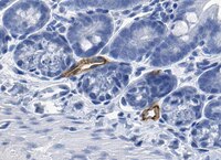
Evaluated by Immunohistochemistry (Paraffin) in mouse colon tissue sections.
Immunohistochemistry (Paraffin) Analysis: A 1:50 dilution of this antibody detected MAdCAM-1 in mouse colon tissue sections.
Usage Statement
Unless otherwise stated in our catalog or other company documentation accompanying the product(s), our products are intended for research use only and are not to be used for any other purpose, which includes but is not limited to, unauthorized commercial uses, in vitro diagnostic uses, ex vivo or in vivo therapeutic uses or any type of consumption or application to humans or animals.
Storage and Shipping Information
Storage Conditions
Stable for 1 year at -20°C from date of receipt.
Handling Recommendations: Upon receipt and prior to removing the cap, centrifuge the vial and gently mix the solution. Aliquot into microcentrifuge tubes and store at -20°C. Avoid repeated freeze/thaw cycles, which may damage IgG and affect product performance.