Wenn Sie das Fenster schließen, wird Ihre Konfiguration nicht gespeichert, es sei denn, Sie haben Ihren Artikel in die Bestellung aufgenommen oder zu Ihren Favoriten hinzugefügt.
Klicken Sie auf OK, um das MILLIPLEX® MAP-Tool zu schließen oder auf Abbrechen, um zu Ihrer Auswahl zurückzukehren.
Wählen Sie konfigurierbare Panels & Premixed-Kits - ODER - Kits für die zelluläre Signaltransduktion & MAPmates™
Konfigurieren Sie Ihre MILLIPLEX® MAP-Kits und lassen sich den Preis anzeigen.
Konfigurierbare Panels & Premixed-Kits
Unser breites Angebot enthält Multiplex-Panels, für die Sie die Analyten auswählen können, die am besten für Ihre Anwendung geeignet sind. Unter einem separaten Register können Sie das Premixed-Cytokin-Format oder ein Singleplex-Kit wählen.
Kits für die zelluläre Signaltransduktion & MAPmates™
Wählen Sie gebrauchsfertige Kits zur Erforschung gesamter Signalwege oder Prozesse. Oder konfigurieren Sie Ihre eigenen Kits mit Singleplex MAPmates™.
Die folgenden MAPmates™ sollten nicht zusammen analysiert werden: -MAPmates™, die einen unterschiedlichen Assaypuffer erfordern. -Phosphospezifische und MAPmate™ Gesamtkombinationen wie Gesamt-GSK3β und Gesamt-GSK3β (Ser 9). -PanTyr und locusspezifische MAPmates™, z.B. Phospho-EGF-Rezeptor und Phospho-STAT1 (Tyr701). -Mehr als 1 Phospho-MAPmate™ für ein einziges Target (Akt, STAT3). -GAPDH und β-Tubulin können nicht mit Kits oder MAPmates™, die panTyr enthalten, analysiert werden.
.
Bestellnummer
Bestellinformationen
St./Pkg.
Liste
Dieser Artikel wurde zu Ihren Favoriten hinzugefügt.
Wählen Sie bitte Spezies, Panelart, Kit oder Probenart
Um Ihr MILLIPLEX® MAP-Kit zu konfigurieren, wählen Sie zunächst eine Spezies, eine Panelart und/oder ein Kit.
Custom Premix Selecting "Custom Premix" option means that all of the beads you have chosen will be premixed in manufacturing before the kit is sent to you.
Catalogue Number
Ordering Description
Qty/Pack
List
Dieser Artikel wurde zu Ihren Favoriten hinzugefügt.
Spezies
Panelart
Gewähltes Kit
Menge
Bestellnummer
Bestellinformationen
St./Pkg.
Listenpreis
96-Well Plate
Menge
Bestellnummer
Bestellinformationen
St./Pkg.
Listenpreis
Weitere Reagenzien hinzufügen (MAPmates erfordern die Verwendung eines Puffer- und Detektionskits)
Menge
Bestellnummer
Bestellinformationen
St./Pkg.
Listenpreis
48-602MAG
Buffer Detection Kit for Magnetic Beads
1 Kit
Platzsparende Option Kunden, die mehrere Kits kaufen, können ihre Multiplex-Assaykomponenten in Kunststoffbeuteln anstelle von Packungen erhalten, um eine kompaktere Lagerung zu ermöglichen.
Dieser Artikel wurde zu Ihren Favoriten hinzugefügt.
Das Produkt wurde in Ihre Bestellung aufgenommen
Sie können nun ein weiteres Kit konfigurieren, ein Premixed-Kit wählen, zur Kasse gehen oder das Bestell-Tool schließen.
Anti-Eosinophil Peroxidase, clone AHE-1, Cat. No. MAB1087-I, is a mouse monoclonal antibody that detects Eosinophil Peroxidase and is tested for use in Immunofluorescence, Immunocytochemistry, Immunohistochemistry.
More>>Anti-Eosinophil Peroxidase, clone AHE-1, Cat. No. MAB1087-I, is a mouse monoclonal antibody that detects Eosinophil Peroxidase and is tested for use in Immunofluorescence, Immunocytochemistry, Immunohistochemistry. Less<<
Empfohlene Produkte
Übersicht
Replacement Information
Description
Catalogue Number
MAB1087-I-25UG
Description
Anti-Eosinophil Peroxidase Antibody, clone AHE-1
Alternate Names
EPO
EPX
Background Information
Eosinophil peroxidase (UniProt: P11678; also known as EC:1.11.1.7, EPO, EPX) is encoded by the EPX (also known as EPER, EPO, EPP) gene (Gene ID: 8288) in human. Eosinophil peroxidase is an enzyme found in cytoplasmic granules of eosinophils. It mediates tyrosine nitration of secondary granule proteins in mature resting eosinophils. It is synthesized with a signal peptide (aa 1-17) and a propeptide (aa 18-139), which are subsequently cleaved off to produce the mature form that can be cleaved into a light chain (aa 140-250) and a heavy chain (aa 251-715). These chains can assemble to for a tetrameric structure. It is a heme-containing enzyme that physically associates with fibrous tissue and cancer tissue in various organs. It contains well-conserved profibrogenic capacity to stimulate the migration of fibroblastic cells and promote their ability to secrete collagenous proteins to generate a functional extracellular matrix (ECM) both in vitro and in vivo. It plays a role in regulating the recruitment of fibroblast and the biosynthesis of collagen ECM at sites of normal tissue repair and fibrosis. It is a key participant in generation of reactive oxidants and diffusible radical species by the phagocytes. It serves as a potent toxin towards invading parasites and the surrounding tissues. It also displays significant inhibitory activity towards Mycobacterium tuberculosis H37Rv by inducing bacterial fragmentation and lysis. (Ref.: DeNichilo, MO., et al. (2015). Am. J. Pathol. 185(5); 1372-1384; Mitra, SN., et al. (2000). Redox Rep. 5(4); 215-224).
References
Product Information
Format
Purified
Presentation
Purified mouse monoclonal antibody IgG1 in buffer containing 0.1 M Tris-Glycine (pH 7.4), 150 mM NaCl with 0.05% sodium azide.
Anti-Eosinophil Peroxidase, clone AHE-1, Cat. No. MAB1087-I, is a mouse monoclonal antibody that detects Eosinophil Peroxidase and is tested for use in Immunofluorescence, Immunocytochemistry, Immunohistochemistry.
Key Applications
Immunofluorescence
Immunohistochemistry
Immunocytochemistry
Application Notes
Tested applications
Immunofluorescence Analysis: A representative lot detected Eosinophil Peroxidase in Immunofluorescence applications (DeNichilo, M.O., et al. (2015). Am J Pathol. 185(5); 1372-84; Song, Y., et al. (2013). ISRN Inflamm. 2013; 907821).
Immunohistochemistry Applications: A representative lot detected Eosinophil Peroxidase in Immunohistochemistry applications (Spang, C., et al. (2017). J Musculoskelet Neuronal Interact. 17(3); 226-236; Song, Y., et al. (2013). ISRN Inflamm. 2013; 907821; Song, Y., et al. (2013). BMC Musculoskelet Disord. 14:; 34; Song, Y., et al. (2012). PLoS One ;7(12); e52230).
Immunocytochemistry Analysis: A representative lot detected Eosinophil Peroxidase in Immunocytochemistry applications (DeNichilo, M.O., et al. (2015). Am J Pathol. 185(5); 1372-84).
Note: Actual optimal working dilutions must be determined by end user as specimens, and experimental conditions may vary with the end user.
Biological Information
Immunogen
Human Eosinophils from a patient with hypereosinophilic syndrome.
Epitope
Unknown
Clone
AHE-1
Concentration
1.0 mg/mL. Please refer to guidance on suggested starting dilutions and/or titers per application and sample type.
Host
Mouse
Specificity
Clone AHE-1 is a mouse monoclonal antibody that detects Eosinophil Peroxidase.
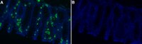
Evaluated by Immunofluorescence in frozen Human colon tissue sections.
Immunofluorescence Analysis: A 1:50 dilution of this antibody detected Eosinophil Peroxidase in frozen Human colon tissue sections.
Usage Statement
Unless otherwise stated in our catalog or other company documentation accompanying the product(s), our products are intended for research use only and are not to be used for any other purpose, which includes but is not limited to, unauthorized commercial uses, in vitro diagnostic uses, ex vivo or in vivo therapeutic uses or any type of consumption or application to humans or animals.