Wenn Sie das Fenster schließen, wird Ihre Konfiguration nicht gespeichert, es sei denn, Sie haben Ihren Artikel in die Bestellung aufgenommen oder zu Ihren Favoriten hinzugefügt.
Klicken Sie auf OK, um das MILLIPLEX® MAP-Tool zu schließen oder auf Abbrechen, um zu Ihrer Auswahl zurückzukehren.
Wählen Sie konfigurierbare Panels & Premixed-Kits - ODER - Kits für die zelluläre Signaltransduktion & MAPmates™
Konfigurieren Sie Ihre MILLIPLEX® MAP-Kits und lassen sich den Preis anzeigen.
Konfigurierbare Panels & Premixed-Kits
Unser breites Angebot enthält Multiplex-Panels, für die Sie die Analyten auswählen können, die am besten für Ihre Anwendung geeignet sind. Unter einem separaten Register können Sie das Premixed-Cytokin-Format oder ein Singleplex-Kit wählen.
Kits für die zelluläre Signaltransduktion & MAPmates™
Wählen Sie gebrauchsfertige Kits zur Erforschung gesamter Signalwege oder Prozesse. Oder konfigurieren Sie Ihre eigenen Kits mit Singleplex MAPmates™.
Die folgenden MAPmates™ sollten nicht zusammen analysiert werden: -MAPmates™, die einen unterschiedlichen Assaypuffer erfordern. -Phosphospezifische und MAPmate™ Gesamtkombinationen wie Gesamt-GSK3β und Gesamt-GSK3β (Ser 9). -PanTyr und locusspezifische MAPmates™, z.B. Phospho-EGF-Rezeptor und Phospho-STAT1 (Tyr701). -Mehr als 1 Phospho-MAPmate™ für ein einziges Target (Akt, STAT3). -GAPDH und β-Tubulin können nicht mit Kits oder MAPmates™, die panTyr enthalten, analysiert werden.
.
Bestellnummer
Bestellinformationen
St./Pkg.
Liste
Dieser Artikel wurde zu Ihren Favoriten hinzugefügt.
Wählen Sie bitte Spezies, Panelart, Kit oder Probenart
Um Ihr MILLIPLEX® MAP-Kit zu konfigurieren, wählen Sie zunächst eine Spezies, eine Panelart und/oder ein Kit.
Custom Premix Selecting "Custom Premix" option means that all of the beads you have chosen will be premixed in manufacturing before the kit is sent to you.
Catalogue Number
Ordering Description
Qty/Pack
List
Dieser Artikel wurde zu Ihren Favoriten hinzugefügt.
Spezies
Panelart
Gewähltes Kit
Menge
Bestellnummer
Bestellinformationen
St./Pkg.
Listenpreis
96-Well Plate
Menge
Bestellnummer
Bestellinformationen
St./Pkg.
Listenpreis
Weitere Reagenzien hinzufügen (MAPmates erfordern die Verwendung eines Puffer- und Detektionskits)
Menge
Bestellnummer
Bestellinformationen
St./Pkg.
Listenpreis
48-602MAG
Buffer Detection Kit for Magnetic Beads
1 Kit
Platzsparende Option Kunden, die mehrere Kits kaufen, können ihre Multiplex-Assaykomponenten in Kunststoffbeuteln anstelle von Packungen erhalten, um eine kompaktere Lagerung zu ermöglichen.
Dieser Artikel wurde zu Ihren Favoriten hinzugefügt.
Das Produkt wurde in Ihre Bestellung aufgenommen
Sie können nun ein weiteres Kit konfigurieren, ein Premixed-Kit wählen, zur Kasse gehen oder das Bestell-Tool schließen.
Anti-DC-STAMP Antibody, clone 1A2 is an antibody against DC-STAMP for use in Western Blotting, Flow Cytometry, Immunocytochemistry, Immunoprecipitation.
More>>Anti-DC-STAMP Antibody, clone 1A2 is an antibody against DC-STAMP for use in Western Blotting, Flow Cytometry, Immunocytochemistry, Immunoprecipitation. Less<<
Anti-DC-STAMP Antibody, clone 1A2 : SDB (Sicherheitsdatenblätter), Analysenzertifikate und Qualitätszertifikate, Dossiers, Broschüren und andere verfügbare Dokumente.
Dendritic cell-specific transmembrane protein (UniProt Q9H295; also known as DC-STAMP, Dendrocyte-expressed seven transmembrane protein, FIND, hDC-STAMP, IL-four-induced protein, Transmembrane 7 superfamily member 4) is encoded by the DCSTAMP (also known as TM7SF4) gene (Gene ID 81501) in human. DC-STAMP is a six-transmembrane protein essential for cell-to-cell fusion to form multinucleated osteoclasts (OCs) during osteoclastogenesis. DC-STAMP expression is upregulated among osteoclast precursor (OCP) cells upon exposure to OC-promoting cytokines, such as receptor activator of nuclear factor-κB (NF-κB) ligand (RANKL), and Dcstamp-knockout (KO) mice have few multinucleated TRAP+ OCs and increased bone mass. On the other hand, DC-STAMP overexpression in transgenic (Tg) mice causes accelerated cell-to-cell fusion during OCP differentiation and enhanced bone resorption. DC-STAMP is a six-trasmembrane (a.a. 35-55, 58-78. 98-118, 210-230, 293-313, 377-397) protein, having both its N- and C-terminal ends exposed intracellularly (a.a. 1-34, 398-470). The C-terminal cytoplasmic tail of DC-STAMP contains an immunoreceptor tyrosine-based inhibitory motif or ITIM sequence (407-SFYPSV-412) that, when phosphorylated on the tyrosine residue, recruits SHP-1. DC-STAMP neutralizing antibody blocks OC formation in vitro and abolishes cellular DC-STAMP and SHP-1 tyrosine phosphorylation.
References
Product Information
Format
Purified
Presentation
Purified mouse monoclonal IgG2aκ antibody in PBS without preservatives.
Anti-DC-STAMP Antibody, clone 1A2 is an antibody against DC-STAMP for use in Western Blotting, Flow Cytometry, Immunocytochemistry, Immunoprecipitation.
Key Applications
Western Blotting
Flow Cytometry
Immunocytochemistry
Immunoprecipitation
Application Notes
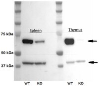
Western Blotting Analysis: 4.0 µg/mL from a representative lot detected DC-STAMP in 10 µg of thymus lysates from wild-type, but not Dcstamp-knockout mice. Western Blotting Analysis: 1.0 µg/mL from a representative lot detected DC-STAMP in thymus and spleen lysates from wild-type mice, and greatly reduced DC-STAMP in thymus and spleen lysates from Dcstamp-knockout mice (Courtesy of Grace Chiu, Ph.D., University of Rochester Medical Center, NY, USA). Western Blotting Analysis: A representative lot detected the ~106 kDa dimeric and the ~53 kDa monomeric DC-STAMP band by Western blotting under non-denatured and denatured condition, respectively, following DC-STAMP immunoprecipitation using murine RAW 264.7 macrophage lysate (Mensah, K.A., et al. (2010). J. Cell. Physiol. 223(1):76-83). Flow Cytometry Analysis: A representative lot detected DC-STAMP-positive lymphoctes and monocytes in purified human PBMCs (Chiu, Y.H., et al. (2012). J. Bone Miner. Res. 27(1):79-92). Flow Cytometry Analysis: A representative lot detected DC-STAMP surface expression on murine RAW 264.7 macrophages and murine bone marrow-derived CD11b+ monocytes. DC-STAMP is expressed on osteoclast precursor (OCP) cells as a dimer, which is efficiently detected by flow cytometry using clone 1A2 (Mensah, K.A., et al. (2010). J. Cell. Physiol. 223(1):76-83). Function Analysis: A representative lot inhibited RANKL & M-CSF treatment-induced osteoclasts (OC) formation in human PBMCs & monocytes cultures (Chiu, Y.H., et al. (2012). J. Bone Miner. Res. 27(1):79-92). Function Analysis: A representative lot inhibited RANKL treatment-induced osteoclasts (OC) formation in RAW 264.7 and murine bone marrow macrophages cultures (Mensah, K.A., et al. (2010). J. Cell. Physiol. 223(1):76-83). Immunohistochemistry Analysis: A representative lot detected DC-STAMP expressionon in multinucleated ‘osteoclast-like’ giant cells in human giant cell tumor of bone (Chiu, Y.H., et al. (2012). J. Bone Miner. Res. 27(1):79-92). Immunocytochemistry Analysis: A representative lot detected DC-STAMP-positive human PBMCs using 10% NBF-fixed, paraffin-embedded cell preparation (Chiu, Y.H., et al. (2012). J. Bone Miner. Res. 27(1):79-92). Immunocytochemistry Analysis: A representative lot detected differential DC-STAMP intracellular localizations by fluorescent immunocytochemistry staining of 4% paraformaldehyde-fixed, 0.1% saponin-permeabilized murine bone marrow macrophages at different time points during osteoclastogenesis upon RANKL treatment (Mensah, K.A., et al. (2010). J. Cell. Physiol. 223(1):76-83). Immunoprecipitation Analysis: A representative lot immunoprecipitated DC-STAMP from the lysates of human monocytes (Chiu, Y.H., et al. (2012). J. Bone Miner. Res. 27(1):79-92). Immunoprecipitation Analysis: A representative lot immunoprecipitated DC-STAMP from the membrane extracts of murine RAW 264.7 macrophages (Mensah, K.A., et al. (2010). J. Cell. Physiol. 223(1):76-83).
Biological Information
Immunogen
KLH-conjugated linear peptide corresponding a sequence from the third extracellular domain of human DC-STAMP.
Epitope
The third extracellular domain.
Clone
1A2
Concentration
Please refer to lot specific datasheet.
Host
Mouse
Specificity
Clone 1A2 recognizes an epitope conserved between human and murine DC-STAMP in the third extracellular domain.
~58 kDa observed. Target band size appears larger than the calculated molecular weights of 53.39 kDa (human) and 53.88 kDa (mouse) due to glycosylation. Uncharacterized band(s) may appear in some lysates.
Physicochemical Information
Dimensions
Materials Information
Toxicological Information
Safety Information according to GHS
Safety Information
Product Usage Statements
Quality Assurance
Evaluated by Western Blotting in mouse thymus lysate.
Western Blotting Analysis: 4.0 µg/mL of this antibody detected DC-STAMP in 10 µg of mouse thymus lysate.
Usage Statement
Unless otherwise stated in our catalog or other company documentation accompanying the product(s), our products are intended for research use only and are not to be used for any other purpose, which includes but is not limited to, unauthorized commercial uses, in vitro diagnostic uses, ex vivo or in vivo therapeutic uses or any type of consumption or application to humans or animals.
Storage and Shipping Information
Storage Conditions
Stable for 1 year at -20°C from date of receipt. Handling Recommendations: Upon receipt and prior to removing the cap, centrifuge the vial and gently mix the solution. Aliquot into microcentrifuge tubes and store at -20°C. Avoid repeated freeze/thaw cycles, which may damage IgG and affect product performance.