Wenn Sie das Fenster schließen, wird Ihre Konfiguration nicht gespeichert, es sei denn, Sie haben Ihren Artikel in die Bestellung aufgenommen oder zu Ihren Favoriten hinzugefügt.
Klicken Sie auf OK, um das MILLIPLEX® MAP-Tool zu schließen oder auf Abbrechen, um zu Ihrer Auswahl zurückzukehren.
Wählen Sie konfigurierbare Panels & Premixed-Kits - ODER - Kits für die zelluläre Signaltransduktion & MAPmates™
Konfigurieren Sie Ihre MILLIPLEX® MAP-Kits und lassen sich den Preis anzeigen.
Konfigurierbare Panels & Premixed-Kits
Unser breites Angebot enthält Multiplex-Panels, für die Sie die Analyten auswählen können, die am besten für Ihre Anwendung geeignet sind. Unter einem separaten Register können Sie das Premixed-Cytokin-Format oder ein Singleplex-Kit wählen.
Kits für die zelluläre Signaltransduktion & MAPmates™
Wählen Sie gebrauchsfertige Kits zur Erforschung gesamter Signalwege oder Prozesse. Oder konfigurieren Sie Ihre eigenen Kits mit Singleplex MAPmates™.
Die folgenden MAPmates™ sollten nicht zusammen analysiert werden: -MAPmates™, die einen unterschiedlichen Assaypuffer erfordern. -Phosphospezifische und MAPmate™ Gesamtkombinationen wie Gesamt-GSK3β und Gesamt-GSK3β (Ser 9). -PanTyr und locusspezifische MAPmates™, z.B. Phospho-EGF-Rezeptor und Phospho-STAT1 (Tyr701). -Mehr als 1 Phospho-MAPmate™ für ein einziges Target (Akt, STAT3). -GAPDH und β-Tubulin können nicht mit Kits oder MAPmates™, die panTyr enthalten, analysiert werden.
.
Bestellnummer
Bestellinformationen
St./Pkg.
Liste
Dieser Artikel wurde zu Ihren Favoriten hinzugefügt.
Wählen Sie bitte Spezies, Panelart, Kit oder Probenart
Um Ihr MILLIPLEX® MAP-Kit zu konfigurieren, wählen Sie zunächst eine Spezies, eine Panelart und/oder ein Kit.
Custom Premix Selecting "Custom Premix" option means that all of the beads you have chosen will be premixed in manufacturing before the kit is sent to you.
Catalogue Number
Ordering Description
Qty/Pack
List
Dieser Artikel wurde zu Ihren Favoriten hinzugefügt.
Spezies
Panelart
Gewähltes Kit
Menge
Bestellnummer
Bestellinformationen
St./Pkg.
Listenpreis
96-Well Plate
Menge
Bestellnummer
Bestellinformationen
St./Pkg.
Listenpreis
Weitere Reagenzien hinzufügen (MAPmates erfordern die Verwendung eines Puffer- und Detektionskits)
Menge
Bestellnummer
Bestellinformationen
St./Pkg.
Listenpreis
48-602MAG
Buffer Detection Kit for Magnetic Beads
1 Kit
Platzsparende Option Kunden, die mehrere Kits kaufen, können ihre Multiplex-Assaykomponenten in Kunststoffbeuteln anstelle von Packungen erhalten, um eine kompaktere Lagerung zu ermöglichen.
Dieser Artikel wurde zu Ihren Favoriten hinzugefügt.
Das Produkt wurde in Ihre Bestellung aufgenommen
Sie können nun ein weiteres Kit konfigurieren, ein Premixed-Kit wählen, zur Kasse gehen oder das Bestell-Tool schließen.
Anti-Cortactin (p80/85), clone 4F11, Cat. No. 05-180-I, is a mouse monoclonal antibody that detects Src substrate protein p85 and is tested for use in Immunocytochemistry, Immunofluorescence, Immunoprecipitation, and Western Blotting.
More>>Anti-Cortactin (p80/85), clone 4F11, Cat. No. 05-180-I, is a mouse monoclonal antibody that detects Src substrate protein p85 and is tested for use in Immunocytochemistry, Immunofluorescence, Immunoprecipitation, and Western Blotting. Less<<
Src substrate protein p85 (UniProt: Q01406; also known as Cortactin, p80) is encoded by the CTTN1 (also known as EMS1, P85.25) gene (Gene ID: 396455) in chicken. Cortactin is an actin filament-binding protein that is localized at cortical regions of cells and serves as a substrate for Src family protein-tyrosine kinases and is shown to be a direct substrate for PER. It is also reported to promote F-actin crosslinking in vitro. Cortactin contributes to the organization of the actin cytoskeleton and cell shape and plays a role in the formation of lamellipodia and in cell migration and regulation of neuron morphology, axon growth and formation of neuronal growth cones. It is also reported to participate in focal adhesion assembly and turnover and intracellular protein transport and endocytosis. In normal cells it is phosphorylated on serine and threonine and in cells expressing activated forms of pp60-src, it becomes heavily phosphorylated on tyrosine residues. Tyrosine phosphorylation of cortactin in transformed cells is shown to contribute to cellular growth regulation and transformation. Cortactin can undergo acetylation by p300 and PCAF acetyltransferase and its acetylation sites are shown to cluster in the repeat region of cortactin harboring its F-actin binding site. Acetylated cortactin loses the positive charge at respective lysines that results in the loss of its F-actin binding. Cortactin contains 7 cortactin repeats and one SH3 motif (aa 505-563). The SH3 motif mediates its binding to the cytoskeleton. Overexpression of cortactin has been reported in B cells from subjects with chronic lymphoblastic leukemia (CLL), however, normal B cells do not express cortactin. (Ref.: Kim, L., and Wong, TW. (1998). J. Biol. Chem. 273(36); 23542-23548; Schnoor, M., et al. (2017). Trends Cell Biol. 28(2); 79-98).
References
Product Information
Format
Purified
Presentation
Purified mouse monoclonal antibody IgG1 in buffer containing 0.1 M Tris-Glycine (pH 7.4), 150 mM NaCl with 0.05% sodium azide.
Anti-Cortactin (p80/85), clone 4F11, Cat. No. 05-180-I, is a mouse monoclonal antibody that detects Src substrate protein p85 and is tested for use in Immunocytochemistry, Immunofluorescence, Immunoprecipitation, and Western Blotting.
Key Applications
Immunocytochemistry
Immunofluorescence
Immunoprecipitation
Western Blotting
Application Notes
Immunocytochemistry Analysis: A representative lot detected Cortactin (p80/85) in Immunocytochemistry applications (Hoffmann, I., et. al. (2001). J Cell Biol. 155(1):133-43; Kim, L., et. al. (1998). J Biol Chem. 273(36):23542-8).
Western Blotting Analysis: A representative lot detected Cortactin (p80/85) in Western Blotting applications (Kim, L., et. al. (1998). J Biol Chem. 273(36):23542-8; Lavoie, J., et. al. (2000). J Cell Biol. 150(5):1037-56; McNiven, M.A., et. al. (2000). J Cell Biol. 151(1):187-98).
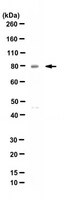
~80 kDa observed; 63.33 and 62.22 kDa, respectively for p85 and p80. Uncharacterized bands may be observed in some lysate(s).
Physicochemical Information
Dimensions
Materials Information
Toxicological Information
Safety Information according to GHS
Safety Information
Product Usage Statements
Quality Assurance
Evaluated by Western Blotting in NIH3T3 cell lysate.
Western Blotting Analysis: A 1:500 dilution of this antibody detected Cortactin (p80/85) in NIH3T3 cell lysate.
Usage Statement
Unless otherwise stated in our catalog or other company documentation accompanying the product(s), our products are intended for research use only and are not to be used for any other purpose, which includes but is not limited to, unauthorized commercial uses, in vitro diagnostic uses, ex vivo or in vivo therapeutic uses or any type of consumption or application to humans or animals.
Storage and Shipping Information
Storage Conditions
Stable for 1 year at +2°C to +8°C from date of receipt.
Activation of ErbB2 receptor tyrosine kinase supports invasion of endothelial cells by Neisseria meningitidis I Hoffmann 1 , E Eugène, X Nassif, P O Couraud, S Bourdoulous J Cell Biol
155(1)
133-43
2001
ErbB2 is a receptor tyrosine kinase belonging to the family of epidermal growth factor (EGF) receptors which is generally involved in cell differentiation, proliferation, and tumor growth, and activated by heterodimerization with the other members of the family. We show here that type IV pilus-mediated adhesion of Neisseria meningitidis onto endothelial cells induces tyrosyl phosphorylation and massive recruitment of ErbB2 underneath the bacterial colonies. However, neither the phosphorylation status nor the cellular localization of the EGF receptors, ErbB3 or ErbB4, were affected in infected cells. ErbB2 phosphorylation induced by N. meningitidis provides docking sites for the kinase src and leads to its subsequent activation. Specific inhibition of either ErbB2 and/or src activity reduces bacterial internalization into endothelial cells without affecting bacteria-induced actin cytoskeleton reorganization or ErbB2 recruitment. Moreover, inhibition of both actin polymerization and the ErbB2/src pathway totally prevents bacterial entry. Altogether, our results provide new insight into ErbB2 function by bringing evidence of a bacteria-induced ErbB2 clustering leading to src kinase phosphorylation and activation. This pathway, in cooperation with the bacteria-induced reorganization of the actin cytoskeleton, is required for the efficient internalization of N. meningitidis into endothelial cells, an essential process enabling this pathogen to cross host cell barriers.
Regulated interactions between dynamin and the actin-binding protein cortactin modulate cell shape M A McNiven 1 , L Kim, E W Krueger, J D Orth, H Cao, T W Wong J Cell Biol
151(1)
187-98
1999
The dynamin family of large GTPases has been implicated in the formation of nascent vesicles in both the endocytic and secretory pathways. It is believed that dynamin interacts with a variety of cellular proteins to constrict membranes. The actin cytoskeleton has also been implicated in altering membrane shape and form during cell migration, endocytosis, and secretion and has been postulated to work synergistically with dynamin and coat proteins in several of these important processes. We have observed that the cytoplasmic distribution of dynamin changes dramatically in fibroblasts that have been stimulated to undergo migration with a motagen/hormone. In quiescent cells, dynamin 2 (Dyn 2) associates predominantly with clathrin-coated vesicles at the plasma membrane and the Golgi apparatus. Upon treatment with PDGF to induce cell migration, dynamin becomes markedly associated with membrane ruffles and lamellipodia. Biochemical and morphological studies using antibodies and GFP-tagged dynamin demonstrate an interaction with cortactin. Cortactin is an actin-binding protein that contains a well defined SH3 domain. Using a variety of biochemical methods we demonstrate that the cortactin-SH3 domain associates with the proline-rich domain (PRD) of dynamin. Functional studies that express wild-type and mutant forms of dynamin and/or cortactin in living cells support these in vitro observations and demonstrate that an increased expression of cortactin leads to a significant recruitment of endogenous or expressed dynamin into the cell ruffle. Further, expression of a cortactin protein lacking the interactive SH3 domain (CortDeltaSH3) significantly reduces dynamin localization to the ruffle. Accordingly, transfected cells expressing Dyn 2 lacking the PRD (Dyn 2(aa)DeltaPRD) sequester little of this protein to the cortactin-rich ruffle. Interestingly, these mutant cells are viable, but display dramatic alterations in morphology. This change in shape appears to be due, in part, to a striking increase in the number of actin stress fibers. These findings provide the first demonstration that dynamin can interact with the actin cytoskeleton to regulate actin reorganization and subsequently cell shape.
Adenovirus E4 open reading frame 4-induced apoptosis involves dysregulation of Src family kinases J N Lavoie 1 , C Champagne, M C Gingras, A Robert J Cell Biol
150(5)
1037-56
1999
The adenoviral early region 4 open reading frame 4 (E4orf4) death factor induces p53-independent apoptosis in many cell types and appears to kill selectively transformed cells. Here we show that expression of E4orf4 in transformed epithelial cells results in early caspase-independent membrane blebbing, associated with changes in the organization of focal adhesions and actin cytoskeleton. Evidence that E4orf4 can associate with and modulate Src family kinase activity, inhibiting Src-dependent phosphorylation of focal adhesion kinase (FAK) and paxillin while increasing phosphorylation of cortactin and some other cellular proteins, is presented. Furthermore, E4orf4 dramatically inhibited the ability of FAK and c-src to cooperate in induction of tyrosine phosphorylation of cellular substrates, suggesting that E4orf4 can interfere with the formation of a signaling complex at focal adhesion sites. Consistent with a functional role for E4orf4-Src interaction, overexpression of activated c-src dramatically potentiated E4orf4-induced membrane blebbing and apoptosis, whereas kinase dead c-src constructs inhibited E4orf4 effects on cell morphology and death. Moreover treatment of E4orf4-expressing cells with PP2, a selective Src kinase inhibitor, led to inhibition of E4orf4-dependent membrane blebbing and later to a marked decrease in E4orf4-induced nuclear condensation. Taken together, these observations indicate that expression of adenovirus 2 E4orf4 can initiate caspase-independent extranuclear manifestations of apoptosis through a modulation of Src family kinases and that these are involved in signaling E4orf4-dependent apoptosis. This study also suggests that Src family kinases are likely to play a role in the cytoplasmic execution of apoptotic programs.
Cortactin associates with the cell-cell junction protein ZO-1 in both Drosophila and mouse T Katsube 1 , M Takahisa, R Ueda, N Hashimoto, M Kobayashi, S Togashi J Biol Chem
273(45)
29672-7
1998
Cortactin is an actin filament-binding protein localizing at cortical regions of cells and a prominent substrate for Src family protein-tyrosine kinases in response to multiple extracellular stimuli. Human cortactin has been identified as a protein product of a putative oncogene, EMS1. In this report, we describe the identification of a Drosophila homolog of cortactin as a molecule that interacts with Drosophila ZO-1 using yeast two-hybrid screening. Drosophila cortactin is a 559-amino acid protein highly expressed in embryos, larvae, and pupae but relatively underexpressed in adult flies. Deletion and substitution mutant analyses revealed that the SH3 domain of Drosophila cortactin binds to a PXXP motif in the proline-rich domain of Drosophila ZO-1. Colocalization of these proteins at cell-cell junction sites was evident under a confocal laser-scanning microscope. In vivo association was confirmed by coimmunoprecipitation of cortactin and ZO-1 from Drosophila embryo lysates. We also demonstrate an association for each of the murine homologs by immunoprecipitation analyses of mouse tissue lysates. Our previous work has demonstrated the involvement of ZO-1 in a signaling pathway that regulates expression of the emc gene in Drosophila. The potential roles of the cortactin.ZO-1 complex in cell adhesion and cell signaling are discussed.
Growth factor-dependent phosphorylation of the actin-binding protein cortactin is mediated by the cytoplasmic tyrosine kinase FER L Kim 1 , T W Wong J Biol Chem
273(36)
23542-8
1998
Previous characterization of the nonreceptor tyrosine kinase FER identified a tight physical association with the catenin pp120 and led to the suggestion that FER may be involved in cell-cell signaling. To further understand the function of FER, we have continued our analyses of the interaction of FER with pp120 and other proteins. The majority of FER is localized to the cytoplasmic fraction where it forms a complex with the actin-binding protein cortactin. The Src homology 2 sequence of FER is required for directly binding cortactin, and phosphorylation of the FER-cortactin complex is up-regulated in cells treated with peptide growth factors. Using a dominant-negative mutant of FER, we provided evidence that FER kinase activity is required for the growth factor-dependent phosphorylation of cortactin. These data suggest that cortactin is likely to be a direct substrate of FER. Our observations provide additional support for a role of FER in mediating signaling from the cell surface, via growth factor receptors, to the cytoskeleton. The nature of the FER-cortactin interaction, and their putative enzyme-substrate relationship, support the previous proposal that one of the functions of the Src homology 2 sequences of nonreceptor tyrosine kinases is to provide a binding site for their preferred substrates.