Wenn Sie das Fenster schließen, wird Ihre Konfiguration nicht gespeichert, es sei denn, Sie haben Ihren Artikel in die Bestellung aufgenommen oder zu Ihren Favoriten hinzugefügt.
Klicken Sie auf OK, um das MILLIPLEX® MAP-Tool zu schließen oder auf Abbrechen, um zu Ihrer Auswahl zurückzukehren.
Wählen Sie konfigurierbare Panels & Premixed-Kits - ODER - Kits für die zelluläre Signaltransduktion & MAPmates™
Konfigurieren Sie Ihre MILLIPLEX® MAP-Kits und lassen sich den Preis anzeigen.
Konfigurierbare Panels & Premixed-Kits
Unser breites Angebot enthält Multiplex-Panels, für die Sie die Analyten auswählen können, die am besten für Ihre Anwendung geeignet sind. Unter einem separaten Register können Sie das Premixed-Cytokin-Format oder ein Singleplex-Kit wählen.
Kits für die zelluläre Signaltransduktion & MAPmates™
Wählen Sie gebrauchsfertige Kits zur Erforschung gesamter Signalwege oder Prozesse. Oder konfigurieren Sie Ihre eigenen Kits mit Singleplex MAPmates™.
Die folgenden MAPmates™ sollten nicht zusammen analysiert werden: -MAPmates™, die einen unterschiedlichen Assaypuffer erfordern. -Phosphospezifische und MAPmate™ Gesamtkombinationen wie Gesamt-GSK3β und Gesamt-GSK3β (Ser 9). -PanTyr und locusspezifische MAPmates™, z.B. Phospho-EGF-Rezeptor und Phospho-STAT1 (Tyr701). -Mehr als 1 Phospho-MAPmate™ für ein einziges Target (Akt, STAT3). -GAPDH und β-Tubulin können nicht mit Kits oder MAPmates™, die panTyr enthalten, analysiert werden.
.
Bestellnummer
Bestellinformationen
St./Pkg.
Liste
Dieser Artikel wurde zu Ihren Favoriten hinzugefügt.
Wählen Sie bitte Spezies, Panelart, Kit oder Probenart
Um Ihr MILLIPLEX® MAP-Kit zu konfigurieren, wählen Sie zunächst eine Spezies, eine Panelart und/oder ein Kit.
Custom Premix Selecting "Custom Premix" option means that all of the beads you have chosen will be premixed in manufacturing before the kit is sent to you.
Catalogue Number
Ordering Description
Qty/Pack
List
Dieser Artikel wurde zu Ihren Favoriten hinzugefügt.
Spezies
Panelart
Gewähltes Kit
Menge
Bestellnummer
Bestellinformationen
St./Pkg.
Listenpreis
96-Well Plate
Menge
Bestellnummer
Bestellinformationen
St./Pkg.
Listenpreis
Weitere Reagenzien hinzufügen (MAPmates erfordern die Verwendung eines Puffer- und Detektionskits)
Menge
Bestellnummer
Bestellinformationen
St./Pkg.
Listenpreis
48-602MAG
Buffer Detection Kit for Magnetic Beads
1 Kit
Platzsparende Option Kunden, die mehrere Kits kaufen, können ihre Multiplex-Assaykomponenten in Kunststoffbeuteln anstelle von Packungen erhalten, um eine kompaktere Lagerung zu ermöglichen.
Dieser Artikel wurde zu Ihren Favoriten hinzugefügt.
Das Produkt wurde in Ihre Bestellung aufgenommen
Sie können nun ein weiteres Kit konfigurieren, ein Premixed-Kit wählen, zur Kasse gehen oder das Bestell-Tool schließen.
Anti-CD34, clone QBEnd/10, Cat. No. CBL496-I, is a mouse monoclonal antibody that detects CD34 and has been tested for use in Electron Microscopy, Flow Cytometry, Immunofluorescence and Fluorescence Activated Cell Sorting (FACS), Immunohistochemistry (Paraffin), and Western Blotting.
More>>Anti-CD34, clone QBEnd/10, Cat. No. CBL496-I, is a mouse monoclonal antibody that detects CD34 and has been tested for use in Electron Microscopy, Flow Cytometry, Immunofluorescence and Fluorescence Activated Cell Sorting (FACS), Immunohistochemistry (Paraffin), and Western Blotting. Less<<
Anti-CD34 Antibody, clone QBEnd/10: SDB (Sicherheitsdatenblätter), Analysenzertifikate und Qualitätszertifikate, Dossiers, Broschüren und andere verfügbare Dokumente.
Hematopoietic progenitor cell antigen CD34 (UniProt: P28906; also known as CD34) is encoded by the CD34 gene (Gene ID: 947) in human. CD34 is a highly glycosylated single-pass type I membrane protein that is expressed on hematopoietic progenitor cells and small vessel endothelium of a variety of tissues. Under normal conditions, CD34+ expressing cells account for about 1 2% of the total bone marrow cells. It serves as an adhesion molecule that plays a role in early hematopoiesis by mediating the attachment of stem cells to the bone marrow extracellular matrix or directly to stromal cells. It is also reported to act as a scaffold for the attachment of lineage specific glycans, allowing stem cells to bind to lectins expressed by stromal cells or other marrow components. CD34 is synthesized with a signal peptide (aa 1-31) that is cleaved off in the mature form. The mature form has an extracellular domain (aa 32-290), a transmembrane domain (aa 291-311), and a cytoplasmic domain (aa 312-385). Two isoforms of CD34 have been described that are produced by alternative splicing.
References
Product Information
Format
Purified
Presentation
Purified mouse monoclonal antibody IgG1 in buffer containing 0.1 M Tris-Glycine (pH 7.4), 150 mM NaCl with 0.05% sodium azide.
Applications
Application
Anti-CD34, clone QBEnd/10, Cat. No. CBL496-I, is a mouse monoclonal antibody that detects CD34 and has been tested for use in Electron Microscopy, Flow Cytometry, Immunofluorescence and Fluorescence Activated Cell Sorting (FACS), Immunohistochemistry (Paraffin), and Western Blotting.
Key Applications
Electron Microscopy
Flow Cytometry
Immunofluorescence
Fluorescence Activated Cell Sorting (FACS)
Immunohistochemistry (Paraffin)
Western Blotting
Application Notes
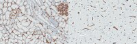
Immunohistochemistry (Paraffin) Analysis: A 1:250 dilution from a representative lot detected CD34 in human brain tissue sections.
Fluorescence Activated Cell Sorting (FACS) Analysis: A representative lot was used to sort CD34+ cells from bone marrow. (de Bock, C.E., et. al. (2012). Leukemia. 26(5):918-26).
Immunofluorescence Analysis: A representative lot detected CD34 in Immunofluorescence applications (Miki, T., et. al. (2010). Mol Cancer Res. 8(5):665-76).
Electron Microscopy Analysis: A representative lot detected CD34 in Electron Microscopy applications (Fina, L., et. al. (1990). Blood. 75(12):2417-26).
Immunohistochemistry Analysis: A representative lot detected CD34 in Immunohistochemistry applications (Engler, J.R., et. al. (2012). PLoS One. 7(8):e43339; Fina, L., et. al. (1990). Blood. 75(12):2417-26).
Flow Cytometry Analysis: A representative lot detected CD34 in Flow Cytometry applications (de Bock, C.E., et. al. (2012). Leukemia. 26(5):918-26; Fina, L., et. al. (1990). Blood. 75(12):2417-26).
Western Blotting Analysis: A representative lot detected CD34 in Western Blotting applications (Fina, L., et. al. (1990). Blood. 75(12):2417-26).
Biological Information
Immunogen
Human placental endothelial membrane vesicles.
Clone
QBEnd/10
Concentration
Please refer to lot specific datasheet.
Host
Mouse
Specificity
Clone QBEnd/10 specifically detects CD34 in human and non-human primates.
40.72 kDa Calculated. This antibody recognizes a heavily glycosylated transmembrane protein: gp 105-120 kDa.
Physicochemical Information
Dimensions
Materials Information
Toxicological Information
Safety Information according to GHS
Safety Information
Product Usage Statements
Quality Assurance
Evaluated by Immunohistochemistry (Paraffin) in human kidney tissue sections.
Immunohistochemistry (Paraffin) Analysis: A 1:250 dilution of this antibody detected CD34 in human kidney tissue sections.
Usage Statement
Unless otherwise stated in our catalog or other company documentation accompanying the product(s), our products are intended for research use only and are not to be used for any other purpose, which includes but is not limited to, unauthorized commercial uses, in vitro diagnostic uses, ex vivo or in vivo therapeutic uses or any type of consumption or application to humans or animals.
Storage and Shipping Information
Storage Conditions
Stable for 1 year at -20°C from date of receipt. Handling Recommendations: Upon receipt and prior to removing the cap, centrifuge the vial and gently mix the solution. Aliquot into microcentrifuge tubes and store at -20°C. Avoid repeated freeze/thaw cycles, which may damage IgG and affect product performance.