Wenn Sie das Fenster schließen, wird Ihre Konfiguration nicht gespeichert, es sei denn, Sie haben Ihren Artikel in die Bestellung aufgenommen oder zu Ihren Favoriten hinzugefügt.
Klicken Sie auf OK, um das MILLIPLEX® MAP-Tool zu schließen oder auf Abbrechen, um zu Ihrer Auswahl zurückzukehren.
Wählen Sie konfigurierbare Panels & Premixed-Kits - ODER - Kits für die zelluläre Signaltransduktion & MAPmates™
Konfigurieren Sie Ihre MILLIPLEX® MAP-Kits und lassen sich den Preis anzeigen.
Konfigurierbare Panels & Premixed-Kits
Unser breites Angebot enthält Multiplex-Panels, für die Sie die Analyten auswählen können, die am besten für Ihre Anwendung geeignet sind. Unter einem separaten Register können Sie das Premixed-Cytokin-Format oder ein Singleplex-Kit wählen.
Kits für die zelluläre Signaltransduktion & MAPmates™
Wählen Sie gebrauchsfertige Kits zur Erforschung gesamter Signalwege oder Prozesse. Oder konfigurieren Sie Ihre eigenen Kits mit Singleplex MAPmates™.
Die folgenden MAPmates™ sollten nicht zusammen analysiert werden: -MAPmates™, die einen unterschiedlichen Assaypuffer erfordern. -Phosphospezifische und MAPmate™ Gesamtkombinationen wie Gesamt-GSK3β und Gesamt-GSK3β (Ser 9). -PanTyr und locusspezifische MAPmates™, z.B. Phospho-EGF-Rezeptor und Phospho-STAT1 (Tyr701). -Mehr als 1 Phospho-MAPmate™ für ein einziges Target (Akt, STAT3). -GAPDH und β-Tubulin können nicht mit Kits oder MAPmates™, die panTyr enthalten, analysiert werden.
.
Bestellnummer
Bestellinformationen
St./Pkg.
Liste
Dieser Artikel wurde zu Ihren Favoriten hinzugefügt.
Wählen Sie bitte Spezies, Panelart, Kit oder Probenart
Um Ihr MILLIPLEX® MAP-Kit zu konfigurieren, wählen Sie zunächst eine Spezies, eine Panelart und/oder ein Kit.
Custom Premix Selecting "Custom Premix" option means that all of the beads you have chosen will be premixed in manufacturing before the kit is sent to you.
Catalogue Number
Ordering Description
Qty/Pack
List
Dieser Artikel wurde zu Ihren Favoriten hinzugefügt.
Spezies
Panelart
Gewähltes Kit
Menge
Bestellnummer
Bestellinformationen
St./Pkg.
Listenpreis
96-Well Plate
Menge
Bestellnummer
Bestellinformationen
St./Pkg.
Listenpreis
Weitere Reagenzien hinzufügen (MAPmates erfordern die Verwendung eines Puffer- und Detektionskits)
Menge
Bestellnummer
Bestellinformationen
St./Pkg.
Listenpreis
48-602MAG
Buffer Detection Kit for Magnetic Beads
1 Kit
Platzsparende Option Kunden, die mehrere Kits kaufen, können ihre Multiplex-Assaykomponenten in Kunststoffbeuteln anstelle von Packungen erhalten, um eine kompaktere Lagerung zu ermöglichen.
Dieser Artikel wurde zu Ihren Favoriten hinzugefügt.
Das Produkt wurde in Ihre Bestellung aufgenommen
Sie können nun ein weiteres Kit konfigurieren, ein Premixed-Kit wählen, zur Kasse gehen oder das Bestell-Tool schließen.
ABC947
Sigma-AldrichAnti-Bif-1
Anti-Bif-1, Cat. No. ABC947, is a rabbit polyclonal antibody that detects Bax-interacting factor 1 (Bif-1) and has been tested for use in Immunohistochemistry (Paraffin) and Western Blotting.
More>>Anti-Bif-1, Cat. No. ABC947, is a rabbit polyclonal antibody that detects Bax-interacting factor 1 (Bif-1) and has been tested for use in Immunohistochemistry (Paraffin) and Western Blotting. Less<<
Anti-Bif-1: SDB (Sicherheitsdatenblätter), Analysenzertifikate und Qualitätszertifikate, Dossiers, Broschüren und andere verfügbare Dokumente.
Endophilin-B1 (UniProt: Q9Y371; also known as Bax-interacting factor 1, Bif-1, SH3 domain-containing GRB2-like protein B1) is encoded by the SH3GLB1 (KIAA0491; CGI-61) gene (Gene ID: 51100) in human. Bif-1 is a member of the endophilin protein family that forms a complex with Beclin-1 through ultraviolet irradiation resistant-associated gene (UVRAG) and promotes the activation of the class III PI3 kinase, Vps34, in mammalian cells. It is highly expressed in heart, skeletal muscle, kidney, and placenta and is detected at lower levels in brain, colon, thymus, spleen, liver, small intestine, lung, and peripheral blood leukocytes. Loss of Bif-1 significantly suppresses PI3KC3 activation and the formation of autophagosome in both HeLa cells and mouse embryonic fibroblasts (MEFs). Bif-1 is reported to be involved in mitochondrial fission and coat protein complex I (COPI)-vesicle formation. Bif-1 contains a BAR domain at the N-terminal (aa 27-261) and a SH3 domain at the C-terminal (305-365), which is required for interaction with UVRAG. Bif-1 may be present as a homodimer or as a heterodimer with Endophilin-B2 (SH3GLB2). Bif-1 can be phosphorylated at Threonine 145 by Cdk5 and this phosphorylation is required for autophagy induction in starved neurons. Three isoforms of Bif-1 have been reported that are generated by alternative splicing. Isoform 1 acts as pro-apoptotic molecule in fibroblasts and is involved in caspase-independent apoptosis during nutrition starvation and in the regulation of autophagy. Isoform 2 has an anti-apoptotic role in neuronal cells and is involved in maintenance of mitochondrial morphology and promotes neuronal viability. (Ref.: Takahashi, Y et al (2009). Cell Death Differen. 16(7), 947-955).
References
Product Information
Format
Affinity Purified
Presentation
Purified rabbit polyclonal antibody in buffer containing 0.1 M Tris-Glycine (pH 7.4), 150 mM NaCl with 0.05% sodium azide.
Anti-Bif-1, Cat. No. ABC947, is a rabbit polyclonal antibody that detects Bax-interacting factor 1 (Bif-1) and has been tested for use in Immunohistochemistry (Paraffin) and Western Blotting.
Key Applications
Immunohistochemistry (Paraffin)
Western Blotting
Application Notes
Western Blotting Analysis: 2 µg/mL from a representative lot detected Bif-1 in 10 µg of Jurkat cells and human kidney lysates.
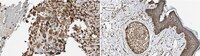
Immunohistochemistry Analysis: A 1:1,000 dilution from a representative lot detected Bif-1 in human placenta, human skin, human small intestine, and human testis tissues.
Biological Information
Immunogen
KLH-conjugated linear peptide corresponding to 15 amino acids from the N-terminal half of human Bax interacting factor 1 (Bif-1).
Concentration
Please refer to lot specific datasheet.
Host
Rabbit
Specificity
This rabbit polyclonal antibody detects Bax interacting facor 1 (Bif-1) in human cells. it targets an epitope within 15 amino acids from the N-terminal region.
~52 kDa observed; 40.80 kDa calculated. Uncharacterized bands may be observed in some lysate(s).
Physicochemical Information
Dimensions
Materials Information
Toxicological Information
Safety Information according to GHS
Safety Information
Product Usage Statements
Quality Assurance
Evaluated by Western Blotting in HEK293 cell lysate.
Western Blotting Analysis: 2 µg/mL of this antibody detected Bif-1 in 10 µg of HEK293 cell lysate.
Usage Statement
Unless otherwise stated in our catalog or other company documentation accompanying the product(s), our products are intended for research use only and are not to be used for any other purpose, which includes but is not limited to, unauthorized commercial uses, in vitro diagnostic uses, ex vivo or in vivo therapeutic uses or any type of consumption or application to humans or animals.