Wenn Sie das Fenster schließen, wird Ihre Konfiguration nicht gespeichert, es sei denn, Sie haben Ihren Artikel in die Bestellung aufgenommen oder zu Ihren Favoriten hinzugefügt.
Klicken Sie auf OK, um das MILLIPLEX® MAP-Tool zu schließen oder auf Abbrechen, um zu Ihrer Auswahl zurückzukehren.
Wählen Sie konfigurierbare Panels & Premixed-Kits - ODER - Kits für die zelluläre Signaltransduktion & MAPmates™
Konfigurieren Sie Ihre MILLIPLEX® MAP-Kits und lassen sich den Preis anzeigen.
Konfigurierbare Panels & Premixed-Kits
Unser breites Angebot enthält Multiplex-Panels, für die Sie die Analyten auswählen können, die am besten für Ihre Anwendung geeignet sind. Unter einem separaten Register können Sie das Premixed-Cytokin-Format oder ein Singleplex-Kit wählen.
Kits für die zelluläre Signaltransduktion & MAPmates™
Wählen Sie gebrauchsfertige Kits zur Erforschung gesamter Signalwege oder Prozesse. Oder konfigurieren Sie Ihre eigenen Kits mit Singleplex MAPmates™.
Die folgenden MAPmates™ sollten nicht zusammen analysiert werden: -MAPmates™, die einen unterschiedlichen Assaypuffer erfordern. -Phosphospezifische und MAPmate™ Gesamtkombinationen wie Gesamt-GSK3β und Gesamt-GSK3β (Ser 9). -PanTyr und locusspezifische MAPmates™, z.B. Phospho-EGF-Rezeptor und Phospho-STAT1 (Tyr701). -Mehr als 1 Phospho-MAPmate™ für ein einziges Target (Akt, STAT3). -GAPDH und β-Tubulin können nicht mit Kits oder MAPmates™, die panTyr enthalten, analysiert werden.
.
Bestellnummer
Bestellinformationen
St./Pkg.
Liste
Dieser Artikel wurde zu Ihren Favoriten hinzugefügt.
Wählen Sie bitte Spezies, Panelart, Kit oder Probenart
Um Ihr MILLIPLEX® MAP-Kit zu konfigurieren, wählen Sie zunächst eine Spezies, eine Panelart und/oder ein Kit.
Custom Premix Selecting "Custom Premix" option means that all of the beads you have chosen will be premixed in manufacturing before the kit is sent to you.
Catalogue Number
Ordering Description
Qty/Pack
List
Dieser Artikel wurde zu Ihren Favoriten hinzugefügt.
Spezies
Panelart
Gewähltes Kit
Menge
Bestellnummer
Bestellinformationen
St./Pkg.
Listenpreis
96-Well Plate
Menge
Bestellnummer
Bestellinformationen
St./Pkg.
Listenpreis
Weitere Reagenzien hinzufügen (MAPmates erfordern die Verwendung eines Puffer- und Detektionskits)
Menge
Bestellnummer
Bestellinformationen
St./Pkg.
Listenpreis
48-602MAG
Buffer Detection Kit for Magnetic Beads
1 Kit
Platzsparende Option Kunden, die mehrere Kits kaufen, können ihre Multiplex-Assaykomponenten in Kunststoffbeuteln anstelle von Packungen erhalten, um eine kompaktere Lagerung zu ermöglichen.
Dieser Artikel wurde zu Ihren Favoriten hinzugefügt.
Das Produkt wurde in Ihre Bestellung aufgenommen
Sie können nun ein weiteres Kit konfigurieren, ein Premixed-Kit wählen, zur Kasse gehen oder das Bestell-Tool schließen.
This Anti-APP Antibody, CT, clone 2.F2.19B4, Ascites Free is validated for use in Immunohistochemistry (Paraffin), Immunocytochemistry, Immunoprecipitation, Western Blotting, Immunofluorescence, Affinity Binding Assay for the detection of APP.
More>>This Anti-APP Antibody, CT, clone 2.F2.19B4, Ascites Free is validated for use in Immunohistochemistry (Paraffin), Immunocytochemistry, Immunoprecipitation, Western Blotting, Immunofluorescence, Affinity Binding Assay for the detection of APP. Less<<
SDB (Sicherheitsdatenblätter), Analysenzertifikate und Qualitätszertifikate, Dossiers, Broschüren und andere verfügbare Dokumente.
Amyloid beta A4 protein (UniProt P05067; also known as ABPP, Alzheimer disease amyloid protein, Amyloid precursor protein, APP, APPI, Cerebral vascular amyloid peptide, CVAP, PN-II, PreA4, Protease nexin-II) is encoded by the APP (also known as A4, AD1) gene (Gene ID 351) in human. Amyloid precursor protein (APP) is initially produced with a signal peptide sequence (a.a. 1-17), the removal of which yields the mature protein with a large extracellular portion (a.a. 18-699), followed by a transmembrane segment (a.a. 700-723) and a cytoplasmic (a.a. 724-770) tail. APP can be further processed by the α-, β-, and γ-secretases in two alternative processing pathways. In the non-amyloidogenic pathway, APP is first cleaved by the plasma membrane-localized α-secretase to generate an N-terminal extracellular sAPPα fragment (a.a. 18-687) and a membrane-bound C-terminal fragment C83 (CTFα), which can be further cleaved by γ-secretase to produce a non-toxic small peptide p3 and a cytoplasmic APP intracellular domain (AICD). In the amyloidogenic pathway, APP undergoes β-cleavage in BACE-1 (β-site APP-cleaving enzyme)-enriched endosomes to generate an N-terminal extracellular sAPPβ fragment (a.a. 18-671) and a membrane-bound C-terminal fragment C99 (CTFβ). Subsequent cleavage of C99 by γ-secretase releases the amyloid β peptides, Aβ1-42 (672-713) & Aβ1-40 (672-711), and AICD. Aβ accumulation in the cortical and hippocampal regions of the brain is a major pathological feature of Alzheimer's disease (AD). Aβ Ser8 phosphorylation is shown to promote Aβ aggregation into oligomeric and fibrillar assemblies and to prevent Aβ proteolytic clearance by certain proteases.
References
Product Information
Format
Purified
Presentation
Purified mouse monoclonal IgG1κ antibody in buffer containing 0.1 M Tris-Glycine (pH 7.4), 150 mM NaCl with 0.05% sodium azide.
This Anti-APP Antibody, CT, clone 2.F2.19B4, Ascites Free is validated for use in Immunohistochemistry (Paraffin), Immunocytochemistry, Immunoprecipitation, Western Blotting, Immunofluorescence, Affinity Binding Assay for the detection of APP.
Key Applications
Immunohistochemistry (Paraffin)
Immunocytochemistry
Immunoprecipitation
Western Blotting
Immunofluorescence
Affinity Binding Assay
Application Notes
Immunocytochemistry Analysis: A representative lot detected a greatly enhanced nuclear APP immunoreactivity due to an upregulated AICD generation and nuclear localization in HSV-1-infected SH-SY5Y cells by fluorescent immunocytochemistry (De Chiara, G., et al. (2010). PLoS One. 5(11):e13989). Immunocytochemistry Analysis: A representative lot immunostained neural progenitor cells (NPCs) isolated from the lateral ventricles of E14 mouse brain by fluorescent immunocytochemistry (Ma, Q.H., et al. (2008). Nat. Cell Biol. 10(3):283-294). Immunoprecipitation Analysis: A representative lot immunoprecipitated APP and cleaved APP C-terminal fragments from uninfected and HSV-1-infected SH-SY5Y cells (De Chiara, G., et al. (2010). PLoS One. 5(11):e13989). Western Blotting Analysis: A representative lot detected APP and cleaved APP C-terminal fragments in lysates and APP immunoprecipitates from uninfected and HSV-1-infected SH-SY5Y cells (De Chiara, G., et al. (2010). PLoS One. 5(11):e13989). Western Blotting Analysis: Representative lots detected the stably expressed full-length human APP695 in HEK293-derived BA-3 cells (Hwang, E.M., et al. (2008). Bioorg. Med. Chem. 16(14):6669-6674; Yeon, S.W., et al. (2007). Peptides. 28(4):838-844). Immunofluorescence Analysis: A representative lot immunostained the walls of the lateral ventricles in E14 mouse brain tissue sections by fluorescent immunohistochemistry (Ma, Q.H., et al. (2008). Nat. Cell Biol. 10(3):283-294). Affinity Binding Assay Analysis: A representative lot captured the synthetic peptide representing APP770 a.a. 732-751 or APP695 a.a. 657-676 (Van Vickle, G.D., et al. (2007). Biochemistry. 46(36):10317-10327).
Biological Information
Immunogen
APP C-terminal 53-a.a. fragment.
Epitope
APP770 a.a. 732-751 (APP695 a.a. 657-676).
Clone
2.F2.19B4
Concentration
Please refer to lot specific datasheet.
Host
Mouse
Specificity
Clone 2.F2.19B4, also known as the Jonas antibody, regcognizes a C-terminal epitope present in 10 of the 11 human spliced isoforms reported by UniProt (P05067), with APP305 being the only spliced isoform lacking the targeted epitope. Clone 2.F2.19B4 recognizes full-length APP, the APP C-terminal fragments (CTFs) produced by α- and β-secretase cleavage, and the APP intracellular domain (AICD) generated by γ-secretase cleavage, but not the sAPPα or sAPPβ fragment. Epitope has been mapped to APP770 a.a. 732-751, which is equivalent to APP695 a.a. 657-676 (Van Vickle, G.D., et al. (2007). Biochemistry. 46(36):10317-10327).
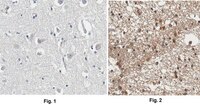
Evaluated by Immunohistochemistry in normal and AD human brain tissue sections.
Immunohistochemistry Analysis: A 1:50 dilution from a representative lot detected APP immunoreactivity in normal human cerebral cortex and Alzheimer's diseased (AD) brain tissue sections.
Usage Statement
Unless otherwise stated in our catalog or other company documentation accompanying the product(s), our products are intended for research use only and are not to be used for any other purpose, which includes but is not limited to, unauthorized commercial uses, in vitro diagnostic uses, ex vivo or in vivo therapeutic uses or any type of consumption or application to humans or animals.