


NA81 Sigma-AldrichAnti-5-Methylcytosine Mouse mAb (162 33 D3)
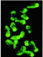

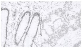
This Anti-5-Methylcytosine Mouse mAb (162 33 D3) is validated for use in ELISA, FC, Frozen Sections, IF, Paraffin Sections, Radioimmunoassay, Southwestern Blot for the detection of 5-Methylcytosine.
More>> This Anti-5-Methylcytosine Mouse mAb (162 33 D3) is validated for use in ELISA, FC, Frozen Sections, IF, Paraffin Sections, Radioimmunoassay, Southwestern Blot for the detection of 5-Methylcytosine. Less<<Anti-5-Methylcytosine Mouse mAb (162 33 D3): SDB (Sicherheitsdatenblätter), Analysenzertifikate und Qualitätszertifikate, Dossiers, Broschüren und andere verfügbare Dokumente.
Synonyme: Anti-5-mc, Anti-5-MeCyd
Empfohlene Produkte
Übersicht
| Replacement Information |
|---|
Key Spec Table
| Species Reactivity | Host | Antibody Type |
|---|---|---|
| A Broad Range Of Species | M | Monoclonal Antibody |
Preis & Verfügbarkeit
| Bestellnummer | Verfügbarkeit | Verpackung | St./Pkg. | Preis | Menge | |
|---|---|---|---|---|---|---|
| NA81-50UG |
|
Kst.-Ampulle | 50 μg |
|
— |

| Product Information | |
|---|---|
| Form | Liquid |
| Formulation | In PBS, pH 7.4. |
| Positive control | NIH3T3 cells |
| Preservative | None |
| Quality Level | MQ100 |
| Physicochemical Information |
|---|
| Dimensions |
|---|
| Materials Information |
|---|
| Toxicological Information |
|---|
| Safety Information according to GHS |
|---|
| Safety Information |
|---|
| Product Usage Statements |
|---|
| Packaging Information |
|---|
| Transport Information |
|---|
| Supplemental Information |
|---|
| Specifications |
|---|
| Global Trade Item Number | |
|---|---|
| Bestellnummer | GTIN |
| NA81-50UG | 04055977209792 |
Documentation
Anti-5-Methylcytosine Mouse mAb (162 33 D3) SDB
| Titel |
|---|
Anti-5-Methylcytosine Mouse mAb (162 33 D3) Analysenzertifikate
| Titel | Chargennummer |
|---|---|
| NA81 |
Literatur
| Übersicht |
|---|
| Ehrlich, M. 2002. Oncogene 21, 5400. (Review) Widschwendter, M. and Jones, P.A. 2002. Oncogene 21, 5462. (Review) Barton, S.C., et al. 2001. Hum. Mol. Genet. 10, 2983. Piyathilake, C.J., et al. 2001. Hum. Pathol. 32, 856. Taddei, A., et al. 2001. Nat. Cell Biol. 3, 114. Piyathilake, C.J., et al. 2000. Biotech. Histochem. 75, 251. De Capoa, A., et al. 1999. FASEB J. 13, 89. |
Broschüre
| Titel |
|---|
| Antibody Sourcebookル Sixth Edition |