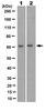
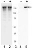
Millipore Sigma Vibrant Logo
Attention: We have moved. Merck Millipore products are no longer available for purchase on MerckMillipore.com.Learn More

CBA073 PhosphoDetect™ c-Met (pTyr1230/1234/1235) ELISA Kit

CBA073
  
Purchase on Sigma-Aldrich

Přehled

Replacement Information

Tabulka spec. kláve

Detection Methods
Colorimetric
Description
Overview

This product has been discontinued.


This product has been discontinued.


We apologize for the inconvenience, but we do not currently have an alternative product.


Detects and quantifies the level of c-Met phosphorylated at Tyr1230/1234/1235. c-Met is a proto-oncogene and a cell-surface receptor for hepatocyte growth factor, which plays an important role in signal transduction process. It is overexpressed in many cancers and often amplified between tumorigenesis and metastasis, so it could be a useful prognostic marker.
Catalogue NumberCBA073
Brand Family Calbiochem®
Application Data
The sensitivity of this ELISA was compared to immunoblotting using known quantities of c-Met (pTyr1230/1234/1235). The data show that the sensitivity of the ELISA is approximately 2x greater than that of immunoblotting. The bands shown in the immunoblot data were developed using an anti-c-Met (pTyr1230/1234/1235) antibody and chemiluminescent detection.
Materials Required but Not Delivered PBS
Plate reader capable of measurement at or near 450 nm
Calibrated adjustable precision pipettes, preferably with disposable plastic tips (a manifold multi-channel pipette is desirable for large assays)
Cell lysis buffer (see Sample Preparation section)
Deionized or distilled H2O
Plate washer: automated or manual (squirt bottle, manifold dispenser, etc.)
Data analysis and graphing software
Glass or plastic tubes for diluting and aliquoting standard
Absorbent paper towels
Calibrated beakers and graduated cylinders in various sizes
References
ReferencesKang, J.Y, et al. 2003. Cancer Res. 63, 1101.
Zhang, X., et al. 2003. Am. J. Renal Physiol. 284, F82.
Maulik, G., et al. 2002. Cytokine Growth Factor Rev. 13, 41.
Crostella, L., et al. 2001. Oncogene 20, 3735.
Fan, S., et al. 2001. Mol. Cell. Biol. 21, 4968.
Longati, P., et al. 2001. Curr. Drug Targets 2, 41.
Matsumoto, K. and Nakamura, T. 2001. Kidney Int. 59, 2023.
Parr, C., et al. 2001. Biochem. Biophys. Res. Commun. 285, 1330.
Follenzi, A., et al. 2000. Oncogene 19, 3041.
Furge, K.A., et al. 2000. Oncogene 19, 5582.
Wei, L., et al. 1995. J. Biol. Chem. 270, 8122.
Guiton, M., et al. 1994. J. Biol. Chem. 269, 30370.
Park, M., et al. 1987. Proc. Natl. Acad. Sci. 84, 6379.
Product Information
Unit of DefinitionOne unit is defined as the amount of c-Met (pTyr<sup>1230/1234/1235</sup>) derived from 21,000 GTL 16 cells treated with 1 mM sodium orthovanadate for 16 h.
Detection methodColorimetric
Form96 Tests
Format96-well plate
Kit containsc-Met (pTyr1230/1234/1235) Standard, Standard Diluent Buffer, c-Met Antibody-Coated 96-Well Plate, Rabbit Anti-c-Met (pTyr1230/1234/1235) Detector Antibody, Anti-Rabbit IgG-HRP Concentrate, HRP Diluent, Wash Buffer Concentrate, TMB, Stop Solution, Plate Covers, and a user protocol.
Positive controlA549 cells
Applications
Biological Information
Assay range1.56-100 Units/ml
Assay time4 h
Sample TypeCells
Physicochemical Information
Sensitivity<0.25 Units/ml
Dimensions
Materials Information
Toxicological Information
Safety Information according to GHS
Safety Information
Product Usage Statements
Intended useThe Calbiochem® PhosphoDetect™ c-Met (pTyr1230/1234/1235) ELISA Kit is designed to detect and quantify the level of c-Met protein that is phosphorylated at Tyr1230/1234/1235. This assay is intended for the detection of c-Met from lysates of human cells. For normalizing c-Met content of the samples, the c-Met ELISA Kit (Cat. No. CBA031), which detects c-Met independent of phosphorylation status, is also available.
Storage and Shipping Information
Ship Code Blue Ice Only
Toxicity Multiple Toxicity Values, refer to MSDS
Storage +2°C to +8°C
Storage ConditionsUpon arrival store the entire contents of the kit at 4°C.
Do not freeze Ok to freeze
Packaging Information
Transport Information
Supplemental Information
Kit containsc-Met (pTyr1230/1234/1235) Standard, Standard Diluent Buffer, c-Met Antibody-Coated 96-Well Plate, Rabbit Anti-c-Met (pTyr1230/1234/1235) Detector Antibody, Anti-Rabbit IgG-HRP Concentrate, HRP Diluent, Wash Buffer Concentrate, TMB, Stop Solution, Plate Covers, and a user protocol.
Specifications
Global Trade Item Number
Katalogové číslo GTIN
CBA073 0

Documentation

PhosphoDetect™ c-Met (pTyr1230/1234/1235) ELISA Kit Certificates of Analysis

TitleLot Number
CBA073

References

Přehled odkazů
Kang, J.Y, et al. 2003. Cancer Res. 63, 1101.
Zhang, X., et al. 2003. Am. J. Renal Physiol. 284, F82.
Maulik, G., et al. 2002. Cytokine Growth Factor Rev. 13, 41.
Crostella, L., et al. 2001. Oncogene 20, 3735.
Fan, S., et al. 2001. Mol. Cell. Biol. 21, 4968.
Longati, P., et al. 2001. Curr. Drug Targets 2, 41.
Matsumoto, K. and Nakamura, T. 2001. Kidney Int. 59, 2023.
Parr, C., et al. 2001. Biochem. Biophys. Res. Commun. 285, 1330.
Follenzi, A., et al. 2000. Oncogene 19, 3041.
Furge, K.A., et al. 2000. Oncogene 19, 5582.
Wei, L., et al. 1995. J. Biol. Chem. 270, 8122.
Guiton, M., et al. 1994. J. Biol. Chem. 269, 30370.
Park, M., et al. 1987. Proc. Natl. Acad. Sci. 84, 6379.

Brochure

Title
Kit SourceBook - 2nd Edition EURO
User Protocol

Revision19-September-2008 RFH
Form96 Tests
Format96-well plate
Detection methodColorimetric
Specieshuman
StorageUpon arrival store the entire contents of the kit at 4°C.
Intended useThe Calbiochem® PhosphoDetect™ c-Met (pTyr1230/1234/1235) ELISA Kit is designed to detect and quantify the level of c-Met protein that is phosphorylated at Tyr1230/1234/1235. This assay is intended for the detection of c-Met from lysates of human cells. For normalizing c-Met content of the samples, the c-Met ELISA Kit (Cat. No. CBA031), which detects c-Met independent of phosphorylation status, is also available.
Backgroundc-Met, a member of the tyrosine kinase superfamily, is the receptor for hepatocyte growth factor, also known as scatter factor (HGF/SF). The mature c-Met protein is a disulfide-linked heterodimer with MW= 190 kDa and composed of a heavily glycosylated α subunit that is completely extracellular in localization, and a β subunit comprised of an extracellular ligand binding domain, a single transmembrane domain, and a cytoplasmic tyrosine kinase domain. c-Met is transcribed from a single open reading frame and translated into a protein precursor that is proteolytically cleaved, yielding the heterodimeric mature protein. Alternative splicing yields several c-Met isoforms, including proteins that remain in the uncleaved, monomeric state or that lack various portions of the c-Met cytoplasmic domain. Cells expressing c-Met include epithelial cells, endothelial cells, blood cells of various types, and glomerular mesenchymal cells. The ligand for c-Met, HGF/SF, is a member of the plasminogen-related growth factor family and is synthesized as an inactive pro-form. HGF/SF activation requires cleavage with either urokinase plasminogen activator (uPA), HGF activator, or Coagulation Factor Xa. Sources of HGF/SF include mesenchymal cells, mesanglial cells, endothelial cells, macrophages, and tumor cells. HGF/SF binding to c-Met stimulates receptor dimerization and the phosphorylation of numerous residues within the receptor's cytoplasmic domain, including Tyr1230/1234/1235 within the Tyr-X-X-X Tyr-Tyr motif of c-Met's activation loop. This motif is conserved among the activation loops of several receptor tyrosine kinases including insulin receptor, insulin like growth factor 1 receptor, nerve growth factor receptor/Trks, and RON. Phosphorylation of Tyr1234/1235 of c-Met is required for activation of the receptor's tyrosine kinase activity. c-Met phosphorylation also generates docking sites for numerous signaling molecules and stimulates receptor internalization via clathrin-coated vesicles. Signaling proteins that are phosphorylated and/or localized in response to c-Met phosphorylation include: Grb2, Shc, Cbl, Crk, cortactin, paxillin, GAB1, PI-3 K, FAK, Src, Ras, ERK1 and 2, JNK, PLC-γ, AKT, and STAT3. HGF/SF stimulation of c-Met expressing cells enhances proliferation, migration, morphogenesis, and protease synthesis, characteristics that are associated with invasive cell phenotype. Indeed, many types of cancer exhibit sustained c-Met stimulation, overexpression, or mutation, including carcinomas of the colon, breast, ovary, lung, liver, prostate, thyroid, kidney, as well as melanomas and sarcomas. In addition to cancer studies, other research areas in which c-Met is under investigation include organogenesis, organ regeneration, angiogenesis, and surgical wound healing.
Principles of the assayThe Calbiochem® c-Met ELISA Kit is a solid phase sandwich Enzyme Linked-Immuno-Sorbent Assay (ELISA). A monoclonal antibody specific for c-Met (regardless of phosphorylation state) has been coated onto the wells of the strips provided. Samples, including a standard containing c-Met, control specimens, and unknowns, are pipetted into these wells. During the first incubation, the c-Met antigen binds to the immobilized (capture) antibody. After washing, a rabbit antibody specific for c-Met phosphorylated at Tyr1230/1234/1235 is added to the wells. During the second incubation, this antibody serves as a detection antibody by binding to the immobilized c-Met protein captured during the first incubation. After removal of excess detection antibody, a horseradish peroxidase-labeled anti-rabbit IgG (anti-rabbit IgG-HRP) is added. This binds to the detection antibody to complete the four-member sandwich. After a third incubation and washing to remove all the excess anti-rabbit IgG-HRP, a substrate solution is added, which is acted upon by the bound enzyme to produce color. The intensity of this colored product is directly proportional to the concentration of c-Met (pTyr1230/1234/1235) present in the original specimen.
Materials provided• c-Met (pTyr1230/1234/1235) Standard (Kit Component No. JA9323-1EA): 2 vials, please refer to the vial label for quantity and reconstitution volume
• Standard Diluent Buffer (Kit Component No. JA9324-25ML): 1 bottle, 25 ml, contains 15 mM sodium azide
• c-Met Antibody-Coated 96-Well Plate (Kit Component No. JA9325-1EA): 1 plate, 96 wells supplied as twelve 8-well strips
• Anti-Rabbit IgG-HRP Concentrate (Kit Component No. JA9327-125UL): 1 vial, 125 µl, supplied as 100X, contains 3.3 mM thymol
• HRP Diluent (Kit Component No. JA9328--25ML): 1 bottle, 25 ml, contains 3.3 mM thymol
• Wash Buffer Concentrate (Kit Component No. JA9329-100ML): 1 bottle, 100 ml, supplied as 25X
• TMB (Kit Component No. JA9330-25ML): 1 bottle, 25 ml, ready-to-use
• Stop Solution (Kit Component No. JA9331-25ML): 1 bottle, 25 ml, ready-to-use
• Plate Cover (Kit Component No. JA9332-1EA): 3 adhesive strips
• Rabbit Anti-c-Met (pTyr1230/1234/1235) Detector Antibody (Kit Component No. JA9326-11ML): 1 bottle, 11 ml, contains 15 mM sodium azide
Materials Required but not provided PBS
Plate reader capable of measurement at or near 450 nm
Calibrated adjustable precision pipettes, preferably with disposable plastic tips (a manifold multi-channel pipette is desirable for large assays)
Cell lysis buffer (see Sample Preparation section)
Deionized or distilled H2O
Plate washer: automated or manual (squirt bottle, manifold dispenser, etc.)
Data analysis and graphing software
Glass or plastic tubes for diluting and aliquoting standard
Absorbent paper towels
Calibrated beakers and graduated cylinders in various sizes
Precautions and recommendations This kit contains materials with small quantities of sodium azide. Sodium azide reacts with lead and copper plumbing to form explosive metal azides. Upon disposal, flush drains with a large volume of water to prevent azide accumulation. Avoid ingestion and contact with eyes, skin and mucous membranes. In case of contact, rinse affected area with plenty of water. Observe all federal, state and local regulations for disposal.
When not in use, kit components should be stored at 4°C. All reagents should be warmed to room temperature before use.
Plates should be allowed to come to room temperature before opening the foil bag. Once the desired number of strips has been removed, immediately reseal the bag and store at 4°C to maintain plate integrity.
Samples should be frozen if not analyzed shortly after collection. Avoid multiple freeze-thaw cycles of frozen samples. Thaw completely and mix well prior to analysis. If large amounts of particulate matter are present, centrifuge or filter prior to analysis.
It is recommended that all standards, controls and samples be run in duplicate.
Samples containing c-Met (pTyr1230/1234/1235) protein should be diluted with Standard Diluent Buffer at least 1:10. This dilution is necessary to reduce the matrix effect of the Cell Lysis Buffer.
When pipetting reagents, maintain a consistent order of addition from well to well. This ensures equal incubation times for all wells.
Cover or cap all reagents when not in use.
• Do not mix or interchange different reagent lots from various kit lots.
Read absorbance within 2 h of assay completion.
In-house controls should be run with every assay. If control values fall outside pre-established ranges, the accuracy of the assay is suspect.
All residual wash liquid must be drained from the wells by efficient aspiration or by decantation followed by tapping the plate forcefully on absorbent paper. Never insert absorbent paper directly into the wells.
Because TMB Substrate is light sensitive, avoid prolonged exposure to light. Also avoid contact between TMB Substrate and metal, or color may develop.
All blood components and biological materials should be handled as potentially hazardous. Follow universal precautions as established by the Centers for Disease Control and Prevention and by the Occupational Safety and Health Administration when handling and disposing infectious agents.
Guidelines for Washing: Incomplete washing will adversely affect the results. All washing must be performed with Wash Buffer Concentrate provided. Washing can be performed manually as follows: completely aspirate the liquid from all wells by gently lowering an aspiration tip (aspiration device) into the bottom of each well. Take care not to scratch the inside of the well. After aspiration, fill the wells with at least 0.4 ml Diluted Wash Buffer. Let soak for 15 to 30 s and aspirate the liquid. Repeat as directed in the Detailed Protocol. Following the final wash, the plate should be inverted and tapped dry on absorbent tissue. Alternatively, the Diluted Wash Buffer may be put into a squirt bottle. If a squirt bottle is used, flood the plate with Diluted Wash Buffer, completely filling all wells. Following the final wash, the plate should be inverted and tapped dry on absorbent tissue. If using an automated washer, the operating instructions for washing equipment should be carefully followed. If your automated washer allows, 30 s soak cycles should be programmed into the wash cycle.
PreparationRecommended Formulation of Cell Lysis Buffer: 10 mM Tris, pH 7.4 100 mM NaCl 1 mM EDTA 1 mM EGTA 1 mM NaF 20 mM Na4P2O7 2 mM Na3VO4 1% Triton® X-100 detergent 10% glycerol 0.1% SDS 0.5% deoxycholate 1 mM PMSF (stock is 0.3 M in DMSO) Protease Inhibitor Cocktail Set III (Cat. No. 539134) This buffer is stable for 2-3 weeks at 4°C or for up to 6 months when aliquoted (without protease inhibitors and PMSF added) and stored at -20°C. When stored frozen, the Cell Lysis Buffer should be thawed on ice. Important: add the protease inhibitors just prior to use. The stability of protease inhibitor supplemented Cell Lysis Buffer is 24 h at 4°C. PMSF is very unstable and must be added prior to use, even if added previously. Protocol for Preparation of Cell Lysates This protocol has been applied to several cell lines using the Cell Lysis Buffer above. Researchers should optimize the cell lysis procedures for their own applications. 1. Collect cells in PBS by centrifugation (non-adherent) or scraping from culture flasks (adherent). 2. Wash cells twice with cold PBS. 3. Remove and discard the supernatant and collect the cell pellet. (At this point the cell pellet can be frozen at -80°C and lysed at a later date). 4. Lyse the cell pellet in Cell Lysis Buffer for 30 min on ice with vortexing at 10 min intervals. The volume of Cell Lysis Buffer depends on the number of cells and expression of c-Met (pTyr1230/1234/1235). For example, 2 x 10⁷ GTL cells grown in RPMI plus 10% FBS and treated with sodium orthovanadate at 1 mM for 16 h can be extracted in 0.5 ml Cell Lysis Buffer. Under these conditions, use of 1-10 µl clarified cell lysate diluted to a volume of 100 µl/well in Standard Diluent Buffer (See Detailed Protocol) is sufficient for the detection of c-Met (pTyr1230/1234/1235). 5. Transfer lysates to microcentrifuge tubes and centrifuge at 13,000 rpm for 10 min at 4°C. 6. Aliquot the clear lysate to clean microcentrifuge tubes. These samples are ready for assay. Lysates can be stored at -80°C. Avoid multiple freeze/thaw cycles.
Reagent preparationNote: This c-Met (pTyr1230/1234/1235) Standard (lyophilized cell extract from GTL 16 cells) is designated in Units/mL. One Unit of standard is equivalent to the amount of c-Met (pTyr1230/1234/1235) derived from ~21,000 GTL 16 cells treated with 1 mM sodium orthovanadate for 16 h. 1. Reconstitute c-Met (pTyr1230/1234/1235) Standard with Standard Diluent Buffer. Refer to the vial label for reconstitution volume and instructions. Swirl or mix gently and then leave the vial for 10 min without mixing to ensure complete reconstitution. Label as 100 Units/ml c-Met (pTyr1230/1234/1235). Use the standard within 1 h of reconstitution. 2. Add 0.25 ml Standard Diluent Buffer to each of 6 tubes labeled 50, 25, 12.5, 6.25, 3.12, and 1.6 Units/ml c-Met (pTyr1230/1234/1235) 3. Make serial dilutions of the Standard as described in the following dilution table. Mix thoroughly between steps. Remaining reconstituted standard should be discarded or frozen at -80°C for further use. Standard can be frozen and thawed one time only without loss of immunoreactivity.


Table 1: Dilution of c-Met (pTyr1230/1234/1235) Standard

• Storage and Final Dilution of Anti-Rabbit IgG Horseradish Peroxidase (HRP) Please Note: The Anti-Rabbit IgG-HRP Concentrate is supplied in 50% glycerol, so the solution is viscous. To ensure accurate dilution, allow the Anti-Rabbit IgG-HRP Concentrate to reach room temperature. Gently mix. Pipette Anti-Rabbit IgG-HRP Concentrate slowly. Remove excess concentrate solution from pipette tip by gently wiping with clean absorbent paper. 1. Add 10 µl Anti-Rabbit IgG-HRP Concentrate to 1 ml HRP Diluent. One ml is sufficient for an 8-well strip. Label as Anti-Rabbit IgG-HRP Working Solution. Use the following guidelines to determine the volume for the number of strips used in the assay.

Table 2: Guidelines for diluting Anti-Rabbit IgG horseradish Peroxidase (HRP)

2. Return the unused Anti-Rabbit IgG-HRP Concentrate to the refrigerator. • Diluted Wash Buffer 1. Allow the Wash Buffer Concentrate reach room temperature and mix to ensure that any precipitated salts are re-dissolved. Dilute 1 volume Wash Buffer Concentrate with 24 volumes deionized water (e.g., 50 ml may be diluted up to 1.25 liters, 100 ml may be diluted up to 2.5 liters). Label as Diluted Wash Buffer. 2. Store both the concentrate and the Diluted Wash Buffer in the refrigerator. The diluted buffer should be used within 14 days.
Detailed protocolBe sure to read the Precautions and Recommendations section before carrying out the assay.

Allow all reagents to reach room temperature before use. Gently mix all liquid reagents prior to use.

Note: A standard curve must be run with each assay.

1. Determine the number of 8-well strips needed for the assay. Insert these in the frame(s) for current use. Return the unused strips to the pouch and store in the refrigerator for future use.
2. Add 100 µl Standard Diluent Buffer to the zero wells. Well(s) reserved for the chromogen blank should be left empty.
3. Add 100 µl standards, samples or controls to the designated wells. Samples prepared in Cell Lysis Buffer must be diluted 1:10 or greater in Standard Diluent Buffer (for example, 10 µl sample into 90 µl buffer). While a 1:10 sample dilution has been found to be satisfactory, higher dilutions such as 1:25 or 1:50 may be optimal. The dilution chosen should be optimized for each experimental system. Tap gently on side of plate to thoroughly mix.
4. Cover the plate with a Plate Cover and incubate for 2 h at room temperature.
5. Thoroughly aspirate or decant the solution from the wells and discard. Wash the wells 4 times. See Guidelines for Washing.
6. Add 100 µl Rabbit Anti-c-Met (pTyr1230/1234/1235) Detector Antibody to each well except the chromogen blank(s). Tap gently on the side of the plate to mix.
7. Cover plate with a Plate Cover and incubate for 1 h at room temperature.
8. Thoroughly aspirate or decant solution from wells and discard the liquid. Wash wells 4 times. See Guidelines for Washing.
9. Add 100 µl Anti-Rabbit IgG-HRP Working Solution to each well except the chromogen blank(s).
10. Cover the plate with the Plate Cover and incubate for 30 min at room temperature.
11. Thoroughly aspirate or decant solution from wells and discard the liquid. Wash wells 4 times. See Directions for Washing.
12. Add 100 µl TMB to each well. The liquid in the wells will begin to turn blue.
13. Incubate for 30 min at room temperature, in the dark. Please Note: Do not cover the plate with aluminum foil or metalized mylar. The incubation time for color development to occur is often determined by the plate reader used. Many plate readers have the capacity to record a maximum absorbance (Abs) of 2.0. The absorbance values should be monitored and the substrate reaction stopped before the absorbance of the positive wells exceeds the limits of the instrument. The absorbance values at 450 nm can only be read after the Stop Solution has been added to each well. If using a reader that records only to 2.0 absorbance points, stopping the assay after 20 to 25 min is suggested.
14. Add 100 µl Stop Solution to each well. Tap side of plate gently to mix. The solution in the wells should change from blue to yellow.
15. Read the absorbance of each well at 450 nm having blanked the plate reader against a chromogen blank composed of 100 µl each TMB and Stop Solution. Read the plate within 2 h of adding the Stop Solution.
16. Plot the absorbance of the standards against the standard concentration. (Optimally, the background absorbance may be subtracted from all data points, including standards, unknowns and controls, prior to plotting.) Draw the best smooth curve through these points to construct the standard curve. If using curve-fitting software, the four-parameter algorithm provides the best curve fit.
17. Read the c-Met (pTyr1230/1234/1235) concentrations for unknown samples and controls from the standard curve plotted in step 16. Multiply value(s) obtained for sample(s) by dilution factor to correct for the dilution in step 3. (Samples still producing signals higher than the highest standard (100 Units/ml) should be further diluted in Standard Diluent Buffer and re-analyzed, multiplying the concentration found by the appropriate dilution factor.)


Example data

Table 3: Example Data

Data obtained for the various standards over the range of 0 to 100 Units/ml c-Met (pTyr1230/1234/1235).

Limitations of the assayDo not extrapolate the standard curve beyond the 100 Units/ml standard point; the dose response is non-linear in this region and accuracy is difficult to obtain. Dilute samples >100 Units/ml with Standard Diluent Buffer; reanalyze these and multiply results by the appropriate dilution factor.
The influence of various lysis buffers has not been thoroughly investigated. The rate of degradation of native c-Met or dephosphorylation of c-Met (pTyr1230/1234/1235) in various matrices has not been investigated. Although CREB degradation or dephosphorylation of c-Met (pTyr1230/1234/1235) in the Cell Lysis Buffer described in this protocol has not been seen to date, the possibility of this occurrence cannot be excluded.
Sensitivity<0.25 Units/ml
Sensitivity NotesThe analytical sensitivity of this assay is <0.25 Units/ml human c-Met (pTyr1230/1234/1235). This was determined by adding two standard deviations to the mean absorbance obtained when the zero standard was assayed 30 times. This level of sensitivity is approximately equivalent to the c-Met (pTyr1230/1234/1235) content of 5,000 cells, lysed as described above.

Figure 1: Sensitivity

The sensitivity of this ELISA was compared to immunoblotting using known quantities of c-Met (pTyr1230/1234/1235). The data show that the sensitivity of the ELISA is approximately 2x greater than that of immunoblotting. The bands shown in the immunoblot data were developed using an anti-c-Met (pTyr1230/1234/1235) antibody and chemiluminescent detection.

Assay Range1.56-100 Units/ml
Precision

Table 4: Intra-Assay Precision

Samples of known c-Met (pTyr1230/1234/1235) concentration were assayed in replicates of 16 to determine precision within an assay.


Table 5: Inter-Assay Precision

Samples were assayed 48 times in multiple assays to determine precision between assays.

RecoveryTo evaluate recovery, Cell Lysis Buffer was diluted 1:10 with Standard Diluent Buffer to bring the SDS concentration to <0.01%. c-Met (pTyr1230/1234/1235) Standard was spiked into the Cell Lysis Buffer. The average recovery was 107%.


Parallelism

Figure 2: Parallelism

Natural c-Met (pTyr1230/1234/1235) from sodium orthovanadate treated GTL 16 and A549 cells was serially diluted in Standard Diluent Buffer. The absorbance of each dilution was plotted against the c-Met (pTyr1230/1234/1235) standard curve. Parallelism was demonstrated by the figure and indicated that the standard accurately reflects c-Met (pTyr1230/1234/1235) content in samples.

Linearity

Table 6: Linearity of Dilution

GTL 16 cells grown in tissue culture medium containing 10% fetal calf serum were treated with sodium orthovanadate at 1 mM for 16 h and lysed with Cell Lysis Buffer. This lysate was diluted in Standard Diluent Buffer over the range of the assay and measured for c-Met (pTyr1230/1234/1235) content. Linear regression analysis of samples versus the expected concentration yielded a correlation coefficient of 0.99.


Specificity

Figure 3: Peptide Blocking

The specificity of this assay for c-Met phosphorylated at Tyr1230/1234/1235 was confirmed by peptide competition. The data presented in Figure 3 show that the phospho-peptide containing the phosphorylated tyrosines could block the ELISA signal. The same c-Met sequences without phosphate group could not block the ELISA signal.


Figure 4: Detection of Phosphorylated and Unphosphorylated c-Met

A549 cells were treated with sodium orthovanadate at 1 mM for 16 hours, lysed, and assayed in parallel for both total c-Met and c-Met (pTyr1230/1234/1235). The figure shows that treatment with sodium orthovanadate, a tyrosine phosphatase inhibitor, increased phosphorylation at 1230, 1234, and 1235 of c-Met in A549 cells.


Figure 5: Specificity

A549 cells were treated with sodium orthovanadate at 1 mM for 16 hours, lysed, and assayed in parallel for both c-Met amount and c-Met (pTyr1230/1234/1235). The amount of c-Met remained comparable, while the levels of phosphorylation at Tyr1230/1234/1235 increased with sodium orthovanadate treatment. The level of c-Met phosphorylation has been normalized for total c-Met content.

Registered TrademarksCalbiochem® is a registered trademark of EMD Biosciences, Inc.
Triton® is a registered trademark of Dow Chemical Company
PhosphoDetect™ and InteractivePathways™ are trademarks of EMD Biosciences, Inc.