Millipore Sigma Vibrant Logo
Attention: We have moved. Merck Millipore products are no longer available for purchase on MerckMillipore.com.Learn More

MABN798 Anti-retSDR1 Antibody, clone A11

MABN798
100 μg  
Purchase on Sigma-Aldrich

Speciální nabídky

Přehled

Replacement Information

Speciální nabídky

Tabulka spec. kláve

Species ReactivityKey ApplicationsHostFormatAntibody Type
H, R, BWB, IH(P), IFMPurifiedMonoclonal Antibody
Description
Catalogue NumberMABN798
DescriptionAnti-retSDR1 Antibody, clone A11
Alternate Names
  • Short-chain dehydrogenase/reductase 3
  • DD83.1
  • Retinol dehydrogenase 17
  • Retinal short-chain dehydrogenase/reductase 1
  • retSDR1
  • Short chain dehydrogenase/reductase family 16C member 1
Background InformationShort-chain dehydrogenase/reductase 3 (EC 1.1.1.300; UniProt O75911; also known as DD83.1, Retinol dehydrogenase 17, Retinal short-chain dehydrogenase/reductase 1, retSDR1, Short chain dehydrogenase/reductase family 16C member 1) is encoded by the DHRS3 (also known as RDH17, SDR16C1) gene (ORF UNQ2424/PRO4983; Gene ID 9249) in human. The retSDR1 enzyme is an SDR (short-chain dehydrogenase/reductase) family member expressed in photoreceptor outer segments, where it mediates the reduction of all-trans-retinal for the regeneration of bleached pigments, the rate limiting step of retinoid reservoir replenishment during the visual cycle. Evidence indicates that retSDR1 also plays a role in tumor suppression. Alternation of vitamin A metabolism seems to be an important event in carcinogenesis and aberrant CpG-island methylation is reported in the DHRS3 promoter region in several neuroblastoma cell lines. The pro-apoptic p53 and p63 are both shown to regulate retSDR1 transcription.
References
Product Information
FormatPurified
PresentationPurified mouse monoclonal IgG1κ antibody in buffer containing 0.1 M Tris-Glycine (pH 7.4), 150 mM NaCl with 0.05% sodium azide.
Quality LevelMQ100
Applications
ApplicationAnti-retSDR1 Antibody, clone A11 is an antibody against retSDR1 for use in Western Blotting, Immunohistochemistry (Paraffin), Immunofluorescence.
Key Applications
  • Western Blotting
  • Immunohistochemistry (Paraffin)
  • Immunofluorescence
Application NotesImmunohistochemistry Analysis: A 1:50 dilution from a representative lot detected retSDR1 immunoreactivity in human and rat retina tissue sections.
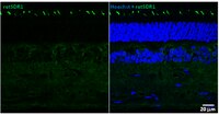
Immunofluorescence Analysis: Clone A11 hybridoma culture supernatant immunostained cone outer segments and a subset of inner nuclear layer somata in 4% paraformaldeyde-fixed, agarose-embedded bovine retina sections by fluorescent confocal microscopy (Courtesy of Francoise Haeseleer, Ph.D., University of Washington, Seattle, WA).
Western Blotting Analysis: A representative lot detected a greatly upregulated retSDR1 protein expression following p53 or TAp63γ overexpression induction in DLD-1 human colorectal adenocarcinoma cells (Kirschner, R.D., et al. (2010). Cell Cycle. 9(11):2177-2188).
Immunofluorescence Analysis: A representative lot immunostained cone, but not rod, outer segments in 4% paraformaldeyde-fixed, agarose-embedded bovine retina sections by fluorescent confocal microscopy. Immunoreactivity was also observed in a subset of somata localized in the inner nuclear layer (Haeseleer, F., et al. (1998). J. Biol. Chem. 273(34):21790-21799).
Immunohistochemistry Analysis: A representative lot immunostained cone, but not rod, outer segments in 4% paraformaldeyde-fixed bovine retina whole mount sections. Immunoreactivity was also observed in a subset of somata localized in the inner nuclear layer (Haeseleer, F., et al. (1998). J. Biol. Chem. 273(34):21790-21799).
Biological Information
ImmunogenHis-tagged recombinant full-length human retSDR1.
EpitopeWithin a.a. 169-303.
CloneA11
ConcentrationPlease refer to lot specific datasheet.
HostMouse
SpecificityClone A11 recognized a truncated human retSDR1 comprising amino acid residues 169–303 by immunoblotting and immunostained cone, but not rod, outer segments by fluorescent confocal microscopy (Haeseleer, F., et al. (1998). J. Biol. Chem. 273(34):21790-21799). Epitope region is absent in human spliced isoform 2 (UniProt O75911-2).
IsotypeIgG1κ
Species Reactivity
  • Human
  • Rat
  • Bovine
Antibody TypeMonoclonal Antibody
Entrez Gene Number
Gene Symbol
  • DHRS3
  • RDH17
  • SDR16C1
  • UNQ2424/PRO4983
Purification MethodProtein G Purified
UniProt Number
Molecular Weight~33 kDa observed. 33.55/33.50/33.61 kDa (human/bovine/rat) calculated. Uncharacterized band(s) may appear in some lysates.
Physicochemical Information
Dimensions
Materials Information
Toxicological Information
Safety Information according to GHS
Safety Information
Product Usage Statements
Quality AssuranceEvaluated by Western Blotting of overexpressed human retSDR1 in HEK293 transfectant lysate.

Western Blotting Analysis: 0.5 µg/mL of this antibody detected the expression of full-length human retSDR1 in 10 µg of lysate from transfected, but not untransfected HEK293 cells.
Usage Statement
  • Unless otherwise stated in our catalog or other company documentation accompanying the product(s), our products are intended for research use only and are not to be used for any other purpose, which includes but is not limited to, unauthorized commercial uses, in vitro diagnostic uses, ex vivo or in vivo therapeutic uses or any type of consumption or application to humans or animals.
Storage and Shipping Information
Storage ConditionsStable for 1 year at 2-8°C from date of receipt.
Packaging Information
Material Size100 μg
Transport Information
Supplemental Information
Specifications
Global Trade Item Number
Katalogové číslo GTIN
MABN798 04055977350364