Disease-associated mutations in Niemann-Pick type C1 alter ER calcium signaling and neuronal plasticity.
Tiscione SA, Vivas O, Ginsburg KS, Bers DM, Ory DS, Santana LF, Dixon RE, Dickson EJ.
J Cell Biol
218(12)
4141-4156
2019
Zobrazit abstrakt
Niemann-Pick type C1 (NPC1) protein is essential for the transport of externally derived cholesterol from lysosomes to other organelles. Deficiency of NPC1 underlies the progressive NPC1 neurodegenerative disorder. Currently, there are no curative therapies for this fatal disease. Given the Ca2+ hypothesis of neurodegeneration, which posits that altered Ca2+ dynamics contribute to neuropathology, we tested if disease mutations in NPC1 alter Ca2+ signaling and neuronal plasticity. We determine that NPC1 inhibition or disease mutations potentiate store-operated Ca2+ entry (SOCE) due to a presenilin 1 (PSEN1)-dependent reduction in ER Ca2+ levels alongside elevated expression of the molecular SOCE components ORAI1 and STIM1. Associated with this dysfunctional Ca2+ signaling is destabilization of neuronal dendritic spines. Knockdown of PSEN1 or inhibition of the SREBP pathway restores Ca2+ homeostasis, corrects differential protein expression, reduces cholesterol accumulation, and rescues spine density. These findings highlight lysosomes as a crucial signaling platform responsible for tuning ER Ca2+ signaling, SOCE, and synaptic architecture in health and disease. | | 31601621
 |
Physical and functional interaction between the α- and γ-secretases: A new model of regulated intramembrane proteolysis.
Chen AC, Kim S, Shepardson N, Patel S, Hong S, Selkoe DJ.
J Cell Biol
211(6)
1157-76
2015
Zobrazit abstrakt
Many single-transmembrane proteins are sequentially cleaved by ectodomain-shedding α-secretases and the γ-secretase complex, a process called regulated intramembrane proteolysis (RIP). These cleavages are thought to be spatially and temporally separate. In contrast, we provide evidence for a hitherto unrecognized multiprotease complex containing both α- and γ-secretase. ADAM10 (A10), the principal neuronal α-secretase, interacted and cofractionated with γ-secretase endogenously in cells and mouse brain. A10 immunoprecipitation yielded γ-secretase proteolytic activity and vice versa. In agreement, superresolution microscopy showed that portions of A10 and γ-secretase colocalize. Moreover, multiple γ-secretase inhibitors significantly increased α-secretase processing (r = -0.86) and decreased β-secretase processing of β-amyloid precursor protein. Select members of the tetraspanin web were important both in the association between A10 and γ-secretase and the γ → α feedback mechanism. Portions of endogenous BACE1 coimmunoprecipitated with γ-secretase but not A10, suggesting that β- and α-secretases can form distinct complexes with γ-secretase. Thus, cells possess large multiprotease complexes capable of sequentially and efficiently processing transmembrane substrates through a spatially coordinated RIP mechanism. | | 26694839
 |
Mint proteins are required for synaptic activity-dependent amyloid precursor protein (APP) trafficking and amyloid β generation.
Sullivan SE, Dillon GM, Sullivan JM, Ho A.
J Biol Chem
289(22)
15374-83
2014
Zobrazit abstrakt
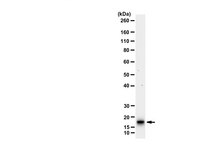
Aberrant amyloid β (Aβ) production plays a causal role in Alzheimer disease pathogenesis. A major cellular pathway for Aβ generation is the activity-dependent endocytosis and proteolytic cleavage of the amyloid precursor protein (APP). However, the molecules controlling activity-dependent APP trafficking in neurons are less defined. Mints are adaptor proteins that directly interact with the endocytic sorting motif of APP and are functionally important in regulating APP endocytosis and Aβ production. We analyzed neuronal cultures from control and Mint knockout neurons that were treated with either glutamate or tetrodotoxin to stimulate an increase or decrease in neuronal activity, respectively. We found that neuronal activation by glutamate increased APP endocytosis, followed by elevated APP insertion into the cell surface, stabilizing APP at the plasma membrane. Conversely, suppression of neuronal activity by tetrodotoxin decreased APP endocytosis and insertion. Interestingly, we found that activity-dependent APP trafficking and Aβ generation were blocked in Mint knockout neurons. We showed that wild-type Mint1 can rescue APP internalization and insertion in Mint knockout neurons. In addition, we found that Mint overexpression increased excitatory synaptic activity and that APP was internalized predominantly to endosomes associated with APP processing. We demonstrated that presenilin 1 (PS1) endocytosis requires interaction with the PDZ domains of Mint1 and that this interaction facilitates activity-dependent colocalization of APP and PS1. These findings demonstrate that Mints are necessary for activity-induced APP and PS1 trafficking and provide insight into the cellular fate of APP in endocytic pathways essential for Aβ production. | Immunoblotting (Western) | 24742670
 |
Role of presenilin 1 in structural plasticity of cortical dendritic spines in vivo.
Jung CK, Fuhrmann M, Honarnejad K, Van Leuven F, Herms J.
J Neurochem
119(5)
1064-73
2010
Zobrazit abstrakt
Mutations in presenilins are the major cause of familial Alzheimer's disease (FAD), leading to impairments of memory and synaptic plasticity followed by age-dependent neurodegeneration. Presenilins are the catalytic subunits of γ-secretase, which itself is critically involved in the processing of amyloid precursor protein to release neurotoxic amyloid β (Aβ). Besides Aβ generation, there is growing evidence that presenilins play an essential role in the formation and maintenance of synapses. To further elucidate the effect of presenilin1 (PS1) on synapses, we performed longitudinal in vivo two-photon imaging of dendritic spines in the somatosensory cortex of transgenic mice over-expressing either human wild-type PS1 or the FAD-mutated variant A246E (FAD-PS1). Interestingly, the consequences of transgene expression were different in two subtypes of cortical dendrites. On apical layer 5 dendrites, we found an enhanced spine density in both mice over-expressing human wild-type presenilin1 and FAD-PS1, whereas on basal layer 3 dendrites only over-expression of FAD-PS1 increased the spine density. Time-lapse imaging revealed no differences in kinetically distinct classes of dendritic spines nor was the shape of spines affected. Although γ-secretase-dependent processing of synapse-relevant proteins seemed to be unaltered, higher expression levels of ryanodine receptors suggest a modified Ca(2+) homeostasis in PS1 over-expressing mice. However, the conditional depletion of PS1 in single cortical neurons had no observable impact on dendritic spines. In consequence, our results favor the view that PS1 influences dendritic spine plasticity in a gain-of-function but γ-secretase-independent manner. | | 21951279
 |


